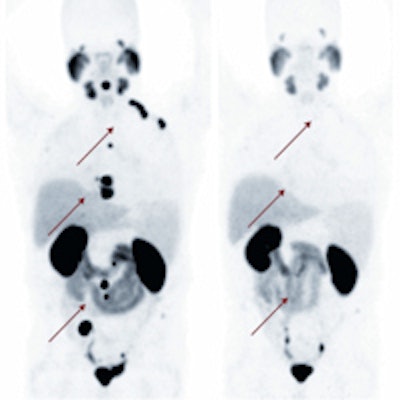
BALTIMORE - German research using a PET radiotracer for prostate cancer that can be labeled with different radioisotopes for diagnostic or therapeutic use won the Image of the Year prize at the Society of Nuclear Medicine and Molecular Imaging (SNMMI) annual meeting.
The emerging imaging technique, developed at Heidelberg University Hospital and the German Cancer Research Center, uses a prostate-specific membrane antigen (PSMA) inhibitor called PSMA-617 to target the enzyme on the surface of prostate cancer cells, even if they have metastasized to other organs. PSMA-617 can be radiolabeled with gallium-68 (Ga-68) for diagnostic use and radiolabeled with lutetium-177 (Lu-177) for therapy.
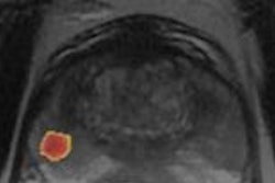
In the study, which was led by Dr. Martina Benesova, follow-up with PET/CT and Ga-68 PSMA-617 showed a substantial reduction in radiotracer uptake, indicating a positive response to therapy. The researchers also found a major decrease in PSMA in the blood, from 38.0 ng/mL to 4.6 ng/mL.
Image courtesy of SNMMI.The preclinical and early clinical experiences with PSMA-617 suggest that it is "suitable for theranostic application due to its high tumor uptake and the highly efficient clearance from the kidneys," wrote Benesova, who is from the division of radiopharmaceutical chemistry at the German Cancer Research Center, and colleagues in a study abstract. "The compound offers a high potential to improve the clinical management of prostate cancer in the future."
In announcing the Image of the Year Award, Dr. Peter Herscovitch, SNMMI's 2014-2015 president, said prostate cancer remains one of the main causes of cancer-related death among men worldwide.
"This new molecular imaging technology not only detects metastatic prostate cancer, but also can treat metastases noninvasively," he added. "It is the combined capability of diagnosis and therapy that makes this molecular theranostic so powerful."
Each year at its annual meeting, SNMMI chooses an image that exemplifies the most promising advances in the field of nuclear medicine and molecular imaging. The technologies captured in the images demonstrate the capacity to improve patient care by detecting disease, aiding diagnosis, improving clinical confidence, and providing a means of selecting appropriate treatments, according to the organization.


![Examples of ultrasound findings and techniques. (A) Images in a 39-year-old male patient with a mass in the left thigh. The mass is heterogeneous on the B-mode US image (compared with the patient in D) and showed increased microvascularity (superb microvascular imaging [SMI]) and shear-wave elastography (SWE) values. Undifferentiated pleomorphic sarcoma was diagnosed at biopsy (with pleomorphic rhabdomyosarcoma in surgical specimen). (B) Images in an 18-year-old male patient with a mass in the left leg. The mass is hypoechoic on the B-mode image, with no other findings suggestive of malignancy. The lesion is in contact with the cortex of the tibia, which is slightly irregular. CT revealed a doubtful anteromedial tibial erosion. The microvascular study demonstrated high vascularization, suggestive of malignancy. Periosteal Ewing sarcoma was diagnosed with both histologic and immunohistochemical confirmation. (C) Images in a 69-year-old female patient with a lump growing on the outside of the left leg. Multiple SWE examinations were performed (please note the high values obtained in the measurements, whereas the color map highlights the stiffness relative to adjacent tissues). SMI showed areas of increased vascularization to target for sampling. Undifferentiated spindle cell sarcoma was diagnosed at biopsy, with residual leiomyosarcoma in the surgical specimen after neoadjuvant therapy. (D) Images in a 56-year-old female patient with a mass in the right thigh. The mass is heterogeneous at both B-mode ultrasound (similar to patient A) and MRI (coronal T2-weighted spectral attenuated inversion recovery [SPAIR]; T1-weighted pre-contrast and postcontrast imaging), which even shows uptake after the administration of paramagnetic contrast material, which is traditionally suggestive of malignancy. Low values at SMI and elastography are suggestive of benignity. Spindle cell lipoma was diagnosed at biopsy, with atypical spindle cell lipomatous tumor in the surgical specimen.](https://img.auntminnieeurope.com/mindful/smg/workspaces/default/uploads/2026/08/images-radiol250278fig2.APCFLSvX6p.jpg?auto=format%2Ccompress&fit=crop&h=100&q=70&w=100)


![Examples of ultrasound findings and techniques. (A) Images in a 39-year-old male patient with a mass in the left thigh. The mass is heterogeneous on the B-mode US image (compared with the patient in D) and showed increased microvascularity (superb microvascular imaging [SMI]) and shear-wave elastography (SWE) values. Undifferentiated pleomorphic sarcoma was diagnosed at biopsy (with pleomorphic rhabdomyosarcoma in surgical specimen). (B) Images in an 18-year-old male patient with a mass in the left leg. The mass is hypoechoic on the B-mode image, with no other findings suggestive of malignancy. The lesion is in contact with the cortex of the tibia, which is slightly irregular. CT revealed a doubtful anteromedial tibial erosion. The microvascular study demonstrated high vascularization, suggestive of malignancy. Periosteal Ewing sarcoma was diagnosed with both histologic and immunohistochemical confirmation. (C) Images in a 69-year-old female patient with a lump growing on the outside of the left leg. Multiple SWE examinations were performed (please note the high values obtained in the measurements, whereas the color map highlights the stiffness relative to adjacent tissues). SMI showed areas of increased vascularization to target for sampling. Undifferentiated spindle cell sarcoma was diagnosed at biopsy, with residual leiomyosarcoma in the surgical specimen after neoadjuvant therapy. (D) Images in a 56-year-old female patient with a mass in the right thigh. The mass is heterogeneous at both B-mode ultrasound (similar to patient A) and MRI (coronal T2-weighted spectral attenuated inversion recovery [SPAIR]; T1-weighted pre-contrast and postcontrast imaging), which even shows uptake after the administration of paramagnetic contrast material, which is traditionally suggestive of malignancy. Low values at SMI and elastography are suggestive of benignity. Spindle cell lipoma was diagnosed at biopsy, with atypical spindle cell lipomatous tumor in the surgical specimen.](https://img.auntminnieeurope.com/mindful/smg/workspaces/default/uploads/2026/08/images-radiol250278fig2.APCFLSvX6p.jpg?auto=format%2Ccompress&fit=crop&h=112&q=70&w=112)