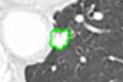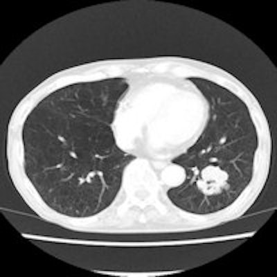
Individuals scanned with CT in the Nederlands-Leuvens Longkanker Screenings Onderzoek (NELSON) lung cancer screening trial had a similar risk of cancer as people who did not participate in the study, meaning the research can be generalized to a wider population.
That's according to a recent analysis of results of the ongoing trial, published in the Journal of Thoracic Oncology (Yousaf-Khan et al, January 30, 2015).
The results suggest that participants in the trial had similar characteristics as the target population for CT lung cancer screening. This is an important consideration when launching a screening program to ensure that disease and mortality rates in actual screening practice will be similar to those predicted by the trial.
The ongoing Dutch-Belgian NELSON trial is investigating whether screening with low-dose CT can reduce lung cancer mortality by at least 25% versus no screening at 10 years of follow-up for individuals 50 to 75 years old. The trial is focusing on individuals with a smoking history of 15 cigarettes or more per day for at least 25 years, or 10 cigarettes or more for at least 30 years. Participants had to be current smokers or have quit 10 or more years ago.
In NELSON, a health questionnaire sent to more than 606,000 individuals ages 50 to 74 years was used to identify at-risk subjects for screening. In all, 15,822 accepted an invitation to screen and were randomized to either low-dose CT or control groups. The remaining individuals were deemed eligible nonparticipants. The current analysis compared the baseline characteristics and mortality profiles of eligible nonparticipants versus the control arm.
Control participants were younger, had better self-reported health, were more physically active, were better educated, and more often were former smokers, compared with the eligible nonparticipants, but numerical differences were minimal, according to the authors. Eligible nonparticipants had higher all-cause mortality as well as mortality due to cardiovascular, respiratory, and noncancerous disease. Even so, the relative proportion of subjects who died due to all types of cancer was higher among participants.
Although eligible nonparticipants were slightly younger and healthier than the screened population, the differences were modest and are unlikely to influence the generalizability of the main results of the trial to the target population, the researchers concluded.


![Examples of ultrasound findings and techniques. (A) Images in a 39-year-old male patient with a mass in the left thigh. The mass is heterogeneous on the B-mode US image (compared with the patient in D) and showed increased microvascularity (superb microvascular imaging [SMI]) and shear-wave elastography (SWE) values. Undifferentiated pleomorphic sarcoma was diagnosed at biopsy (with pleomorphic rhabdomyosarcoma in surgical specimen). (B) Images in an 18-year-old male patient with a mass in the left leg. The mass is hypoechoic on the B-mode image, with no other findings suggestive of malignancy. The lesion is in contact with the cortex of the tibia, which is slightly irregular. CT revealed a doubtful anteromedial tibial erosion. The microvascular study demonstrated high vascularization, suggestive of malignancy. Periosteal Ewing sarcoma was diagnosed with both histologic and immunohistochemical confirmation. (C) Images in a 69-year-old female patient with a lump growing on the outside of the left leg. Multiple SWE examinations were performed (please note the high values obtained in the measurements, whereas the color map highlights the stiffness relative to adjacent tissues). SMI showed areas of increased vascularization to target for sampling. Undifferentiated spindle cell sarcoma was diagnosed at biopsy, with residual leiomyosarcoma in the surgical specimen after neoadjuvant therapy. (D) Images in a 56-year-old female patient with a mass in the right thigh. The mass is heterogeneous at both B-mode ultrasound (similar to patient A) and MRI (coronal T2-weighted spectral attenuated inversion recovery [SPAIR]; T1-weighted pre-contrast and postcontrast imaging), which even shows uptake after the administration of paramagnetic contrast material, which is traditionally suggestive of malignancy. Low values at SMI and elastography are suggestive of benignity. Spindle cell lipoma was diagnosed at biopsy, with atypical spindle cell lipomatous tumor in the surgical specimen.](https://img.auntminnieeurope.com/mindful/smg/workspaces/default/uploads/2026/08/images-radiol250278fig2.APCFLSvX6p.jpg?auto=format%2Ccompress&fit=crop&h=100&q=70&w=100)


![Examples of ultrasound findings and techniques. (A) Images in a 39-year-old male patient with a mass in the left thigh. The mass is heterogeneous on the B-mode US image (compared with the patient in D) and showed increased microvascularity (superb microvascular imaging [SMI]) and shear-wave elastography (SWE) values. Undifferentiated pleomorphic sarcoma was diagnosed at biopsy (with pleomorphic rhabdomyosarcoma in surgical specimen). (B) Images in an 18-year-old male patient with a mass in the left leg. The mass is hypoechoic on the B-mode image, with no other findings suggestive of malignancy. The lesion is in contact with the cortex of the tibia, which is slightly irregular. CT revealed a doubtful anteromedial tibial erosion. The microvascular study demonstrated high vascularization, suggestive of malignancy. Periosteal Ewing sarcoma was diagnosed with both histologic and immunohistochemical confirmation. (C) Images in a 69-year-old female patient with a lump growing on the outside of the left leg. Multiple SWE examinations were performed (please note the high values obtained in the measurements, whereas the color map highlights the stiffness relative to adjacent tissues). SMI showed areas of increased vascularization to target for sampling. Undifferentiated spindle cell sarcoma was diagnosed at biopsy, with residual leiomyosarcoma in the surgical specimen after neoadjuvant therapy. (D) Images in a 56-year-old female patient with a mass in the right thigh. The mass is heterogeneous at both B-mode ultrasound (similar to patient A) and MRI (coronal T2-weighted spectral attenuated inversion recovery [SPAIR]; T1-weighted pre-contrast and postcontrast imaging), which even shows uptake after the administration of paramagnetic contrast material, which is traditionally suggestive of malignancy. Low values at SMI and elastography are suggestive of benignity. Spindle cell lipoma was diagnosed at biopsy, with atypical spindle cell lipomatous tumor in the surgical specimen.](https://img.auntminnieeurope.com/mindful/smg/workspaces/default/uploads/2026/08/images-radiol250278fig2.APCFLSvX6p.jpg?auto=format%2Ccompress&fit=crop&h=112&q=70&w=112)