Doctors are changing the way they examine and diagnose their patients, thanks to the advent of less expensive, portable ultrasound scanners, according to a 29 May article in BMJ.
Dr. Marc Wittenberg, a clinical fellow with the National Health Service England and an editor of BMJ, wrote that general practitioners and specialists "are increasingly using ultrasound to make on-the-spot diagnoses of many conditions without having to consult an imaging expert."
Ultrasound has also become more commonly used because the modality does not expose patients to ionizing radiation, as they normally would be with a CT scan or an x-ray.
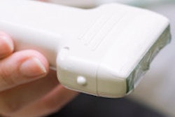
The latest portable machines produce images that are almost the same quality as that of the larger scanners, and they are easy to use, durable, and cost as little as 5,000 pounds (6,160 euros). The result is that doctors in all sorts of fields are starting to use them and, with a growing body of literature supporting their use in the developing world, the World Health Organization now recommends them as a primary diagnostic tool in low-resource environments.
Wittenberg also cited a report that found the number of ultrasound examinations performed by imaging professionals increased on average by 5.2% every year for the past 10 years.


![Overview of the study design. (A) The fully automated deep learning framework was developed to estimate body composition (BC) (defined as subcutaneous adipose tissue [SAT] in liters; visceral adipose tissue [VAT] in liters; skeletal muscle [SM] in liters; SM fat fraction [SMFF] as a percentage; and intramuscular adipose tissue [IMAT] in deciliters) from MRI. The fully automated framework comprised one model (model 1) to quantify different BC measures (SAT, VAT, SM, SMFF, and IMAT) as three-dimensional (3D) measures from whole-body MRI scans. The second model (model 2) was trained to identify standardized anatomic landmarks along the craniocaudal body axis (z coordinate field), which allowed for subdividing the whole-body measures into different subregions typically examined on clinical routine MRI scans (chest, abdomen, and pelvis). (B) BC was quantified from whole-body MRI in over 66,000 individuals from two large population-based cohort studies, the UK Biobank (UKB) (36,317 individuals) and the German National Cohort (NAKO) (30,291 individuals). Bar graphs show age distribution by sex and cohort. BMI = body mass index. (C) After the performance assessment of the fully automated framework, the change in BC measures, distributions, and profiles across age decades were investigated. Age-, sex-, and height-adjusted body composition reference curves were calculated and made publicly available in a web-based z-score calculator (https://circ-ml.github.io).](https://img.auntminnieeurope.com/mindful/smg/workspaces/default/uploads/2026/05/body-comp.XgAjTfPj1W.jpg?auto=format%2Ccompress&fit=crop&h=112&q=70&w=112)


