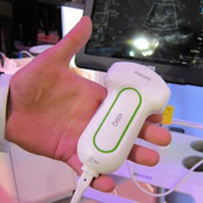
Philips Healthcare is entering the compact ultrasound segment with the launch in India of its new Visiq handheld scanner.
Visiq is designed to be taken wherever healthcare is needed, such as labor rooms and remote areas, for ob/gyn and abdominal scanning, according to the company. The system can scan for 2.5 hours on a single battery charge, and wakes from sleep mode to be scan-ready in seconds.
 The new Philips Visiq scanner.
Visiq's touchscreen controls are similar to those of a smartphone or tablet, and users can capture images, acquire measurements, and share data over built-in Wi-Fi for DICOM data transfer. The scanner also includes many of the same image optimization protocols on the company's premium Epiq ultrasound scanners.
The scanner includes a broadband digital beamformer and sophisticated image acquisition module, with 2D grayscale pulse Doppler and color Doppler modes.
Philips introduced the scanner on 2 April at the Philips Innovation Summit in Bangalore; the system is not yet available in the U.S.
Philips used the summit to highlight a number of other technologies developed specifically for South Asia and other growth regions. In healthcare, these included a mobile obstetrical monitoring solution designed to support telehealth-based detection of high-risk pregnancies in regions where maternal mortality is a problem. Midwives using mobile phones can collect data from physical exams and transmit it to ob/gyn physicians in different locations.
The company also discussed IntelliSpace Consultative Critical Care, telehealth software for remote intensive care delivery units (ICU) in rural and urban areas of India where specialist care is not always available. Patients with serious injuries or illness can be monitored by ICU specialists from a central hospital using audiovisual technology.