Simultaneous PET/MRI has the potential to improve patient care, reduce hospital visits, and provide new and more accurate markers for disease assessment, according to University College London researchers. Early proof-of-concept studies show the clinical performance of PET/MRI is on par with PET/CT.
Interest has been growing in the combination of PET/MRI due to the power of MRI for tissue characterization, microstructural appraisal, and functional assessment, together with new PET tracers designed to target specific metabolic processes. As a result, software-based fusion of images acquired with separate PET and MRI machines has been most common, but recently manufacturers have integrated PET and MRI hardware into a single system that acquires images from both modalities simultaneously, the U.K. researchers explained (British Journal of Radiology [BJR], 14 November 2013).

 Left: Apparent diffusion coefficient (ADC) map derived from b0 - b800 diffusion-weighted images corresponding to the same patient demonstrates a hindered ADC within the left cervical lymph nodes in keeping with elevated cellularity. Right: Contrast-inverted b800 diffusion-weighted image of the same patient as illustrated left. Dark areas indicate increased diffusion signal within the involved left cervical lymph nodes. All images courtesy of Dr. Shonit Punwani.
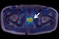
 Left: Axial F-18 FDG PET uptake in the same patient is illustrated within the left cervical lymph node in keeping with increased glucose metabolic activity. Right: Coronal Dixon T1-weighted in-phase MR image with superimposed F-18 FDG PET uptake. Left cervical lymphadenopathy is demonstrated in a patient with a known diagnosis of Hodgkin's lymphoma.
One of the first commercially available PET/MRI systems is from Siemens Healthcare, Biograph mMR. The scanner has a 60-cm bore and 3-tesla magnet that integrates a ring of solid-state PET detectors that are insensitive to magnetic fields, enabling the system to simultaneously acquire PET and MRI signals.
Drs. Francesco Fraioli, a nuclear medicine physician from the Institute of Nuclear Medicine, and Shonit Punwani, a radiologist from the Center for Medical Imaging at University College London, reviewed the potential clinical and research application of simultaneous PET/MRI for assessment of neurological, body oncological, cardiovascular, and inflammatory diseases.
"The next step will be to generate initial clinical applications together with optimized protocols for subsequent validation and evaluation of clinical benefit," they wrote. "Much work remains to be done before the techniques can become part of a standard clinical diagnostic pathway."
Applying PET/MRI
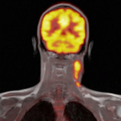
Brain imaging was one of the first applications for simultaneous PET/MRI systems because the brain is less prone to patient motion. What's more, interest is growing within the neurological community for novel radiopharmaceutical tracers to evaluate neurodegenerative and oncological disease with PET/MRI.
PET/MRI is particularly useful in imaging patients with dementia and Alzheimer's disease, because the combination of high-resolution anatomical, perfusion, and tractography MRI information with F-18 FDG or Pittsburgh Compound B (PiB) PET during a single simultaneous examination creates opportunities for improving the understanding of pathogenesis and mechanism of Alzheimer's disease, and enables early diagnosis and supports drug development.
Epilepsy is another condition PET/MRI can help with. A match between areas of hypometabolism and the epileptogenic focus has been demonstrated on interictal PET. Moreover, preliminary results show a possible mismatch of metabolism and perfusion.
"It is worth highlighting that in this disease there maybe discrepancies in either metabolism and/or tissue microstructure and/or vascularity, making the possible use of a combined simultaneous PET/MRI technique important for diagnosis and treatment planning," Fraioli and colleagues wrote.
For intracranial neoplasms, a PET/MRI method combining gadolinium enhancement and somatostatin tracer uptake could have the potential to further improve preoperative planning and better monitor and localize recurrence following therapy.
Body oncological applications
PET/CT is the typical imaging modality for cancer staging, but the noncontrast-enhanced CT provides little tissue characterization beyond the assessment of density. While multiparametric MRI has emerged as the method of choice, it remains more difficult to assess metabolic activity with MRI, and MRI signals themselves are not as specific as PET tracers. Simultaneous PET/MRI combines the power of MRI tissue characterization with targeted molecular PET imaging, as well as reduces both spatial and temporal correlation errors.
For lymphoma, increased glucose metabolism is common to most lymphoma subtypes, and PET with F-18 FDG has become an established method to stage lymphoma; a recent study demonstrated the feasibility of F-18 FDG PET/MRI for lymphoma treatment response evaluation, and the researchers reported good image quality of the PET/MRI examination and an excellent interobserver agreement for Ann Arbor stage, which Fraioli and colleagues' experience corroborates (Magnetic Resonance Materials in Physics, Biology and Medicine, February 2013, Vol. 26:1, pp. 49-55).
PET/MRI is also useful in prostate cancer, head and neck squamous cell carcinoma, neuroendocrine tumors, gastrointestinal tumors, gynecological tumors, and musculoskeletal tumors.
Cardiovascular applications
"PET/MRI for assessment of cardiovascular disease may truly exploit and require simultaneous acquisition of PET and MRI signals," the researchers wrote. "PET is the gold standard for noninvasive assessment of myocardial viability. For the first time, simultaneous PET/MRI can provide direct comparison of the diagnostic performance of each modality at identical resting and stress conditions."
Also, there is the potential for morphologic information obtained by MRI to be combined with PET functional imaging --- for instance, to help differentiate between epicardial stenosis and microvascular dysfunction, or between a scar and dysfunctional but viable myocardium.
PET/MRI can also follow molecular and cellular events after myocardial infarction.
Inflammatory disease applications
Advances in multidetector-row CT and MRI pulse sequences have improved anatomical detail and tissue characterization of inflammatory diseases; several efforts have been made to combine new MR contrast agents with PET imaging.
"There is an interest in developing and assessing MRI contrast agents labeled with PET tracer, where MR contrast agents are used to provide high-resolution anatomical detail of the location of accumulated metabolic PET tracer activity within plaques," the authors wrote.
PET/MRI could also be used in the assessment of inflammatory bowel disease as the hybrid modality could determine if metabolic PET information can complement MRI and thereby guide therapy.
The researchers caution that more research needs to be done before PET/MRI becomes standard clinical practice for any of the aforementioned conditions.