We don't necessarily think high fashion when picturing radiology -- more like lab coats and lead aprons. But London-based radiographer-turned-designer Brooke Roberts has bridged the two worlds with her clothing line that draws upon medical imaging for inspiration.
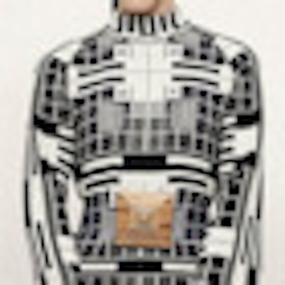
 Roberts' "Calibration" line draws inspiration from x-ray calibration films. Image credit: Philip Meech.
Her knitwear collections are influenced by everything from the different opacities of CT scans to the geometry of x-ray calibration films, often with a focus on brain imaging, which helped lead her from medical imaging to fashion.
"I was performing a CT brain scan and wondered how -- and if -- it could translate to knit," she said. "So I began the experiment at a factory in Italy, programming [knitting machines to create] knitwear from brain scans."
Roberts has a degree in applied science and has worked as a radiographer at several London hospitals, including the North Middlesex, the Heart Hospital, and the London Independent. She hasn't cast aside medical imaging for fashion, though, and continues to work as a radiographer, focusing on cardiac angiography for the past eight years. "I also love CT because I find the images so inspiring," she added.
To transition toward design, she studied at London College of Fashion and Central Saint Martins, while still working as a radiographer. There she was drawn to the pattern-making part of the design process because it involves both creative and technical skills.
 Radiographer and designer Brooke Roberts.
"Then I grew into a knitwear designer," she said. "I work with digital knit techniques, programming and experimenting widely, which is where my applied science background helps."
To Roberts, working in both radiology and fashion seems quite natural. "As Einstein said: Religion, art, and science are all branches of the same tree," she noted. "Art is about asking questions; science is about providing answers. They're perfect bedfellows. Both enrich and engage the imagination."
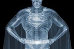
She looked to both x-ray and CT for inspiration when creating her spring/summer 2011 "Cuts" collection, which reflects the transparency and opacity of anatomy in images.
"I used ... mixed transparent and opaque yarns to reflect the absorption of denser tissue and the radiolucency of air seen on x-ray in the form of a knitted textile," she said. With brain CT scans in mind, she paired a fine, semitransparent yarn with an opaque one to create a double-layered fabric.
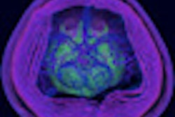
Brain imaging also led Roberts in developing a 2012 line based on scans from the Allen Institute for Brain Science, a nonprofit organization in Seattle dedicated to brain research. In 2011, she received a Creatives in Residence award from the Hospital Club, a London group promoting the arts. The club and the Allen Institute are both supported by Microsoft co-founder Paul Allen.
 Examples of the "Cuts" collection, which is based on CT and x-ray scans. Image credit: Philip Meech.
"Since I presented the collection at the club, Jody Allen, Paul Allen's sister, has been a supporter of my work, which is wonderful," she said. Jody Allen co-founded the brain institute with her brother.
"Brain imaging has fascinated me since I began studying CT, and the Allen Institute images were particularly abstract, which made for interesting artwork," she said. "Translating them into knit was a new challenge, which opened my eyes to other forms of medical imaging."
 The collection based on the Allen Institute for Brain Science images was presented during London Fashion Week. Image credit: Philip Meech.
In the future, Roberts would potentially like to explore embroidered surgical devices -- essentially, devices such as stents or implants created using embroidery -- and the technology used to produce them. She is also interested in eventually expanding to men's clothing.
Her current collections can be viewed at BrookeRoberts.net. The cost of each piece ranges from approximately 200 to 400 pounds (250 to 500 euros).