It seems that art and science do not always mix, at least according to the French Nuclear Safety Authority (ASN). Triggered by an art exhibit involving roughly 100 hand radiographs, the agency served a reminder to those involved and the general public that x-ray scans for artistic purposes are considered unlawful.
ASN was notified at the end of 2012 about artist Marc Ferrante's "Jeux de mains" or Hand Plays project, which was on display at locations in Strasbourg and Maxéville, according to a statement on the ASN site. Between 6 February and 23 April 2013, the Strasbourg division of the agency contacted the artist and inspected three imaging facilities involved in obtaining the x-ray scans.
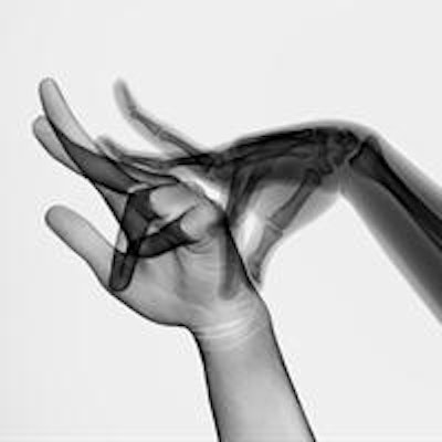
 "Skin and bones" from the Hand Plays modified presence series. All images © Marc Ferrante (marcferrante.com).
The impetus behind the investigation was Article L. 1333-11 of the Public Health Code, which states that ionizing radiation cannot be used on the human body for purposes other than diagnosis, treatment, or biomedical research. The penalty for infringement can range up to one year in prison and a fine of 15,000 euros, ASN noted.
The Hand Plays project is divided into six series of images. In the "object theater" series, for example, scans were taken of hands wearing a range of objects, from more traditional puppets to poultry. Other series include "skin," in which the bones are removed, leaving only an image of the skin, and "ergon," which visualizes the space between the hands.
The collection aims to examine radiography in light of its many influences, according to Ferrante. "Like any other type of images, x-rays are loaded with historical and cultural references that inform and change our way of looking at imagery, without us necessarily being aware of it," he said.
 "Rabbit" from the shadow play series.
Ferrante worked with a variety of individuals on Hand Plays, from radiologists, radiographers, and surgeons to puppeteers, marionettists, and dancers. The project also included support from Fujifilm Medical Systems, BSN Medical, and various organizations.
Inspections
According to ASN, approximately 25 volunteers were scanned at the three imaging sites: Centre hospitalier universitaire (CHU) de Hautepierre in Strasbourg, Cabinet de radiologie in Strasbourg, and SEL d'imagerie médicale "Majorelle" in Nancy. The inspections concluded that the doses administered were not enough to significantly affect the volunteers' health; however, the agency also warned that such imaging must not be conducted without proper cause.
Ferrante said he felt the images didn't put anyone in danger. "This is the reason why radiologists and manufacturers have helped me; this is also the reason why ASN confined itself to a [reminder] of the law," he told AuntMinnieEurope.com.
Letters to the three sites summarizing the inspections were published on the ASN website for the public. Based on the results of the inspections, the groups were asked to ensure compliance with L.1333-11, as well as provide the agency with documentation confirming that all relevant professionals have undergone the appropriate training regarding radiation safety for patients.
Ferrante said he does understand the concern and the regulation, especially in a historical context.
"Radiography had many victims at the beginning of the 20th century, especially among doctors, because at the time the x-ray doses were extremely high, and it took quite a while before people started protecting themselves correctly," he said. "The current regulations are therefore logical consequences."
"Today, even though the use of x-rays is still dangerous, the doses are a lot less significant, often miniscule, and for the most part they have much less of an impact on our health thanks to the digitization of the radiographic process," he noted.
 Hand Plays is currently on display at the Muséum-Aquarium of Nancy.
Ferrante has spent several years investigating the technology but intends to move on to other areas for future work.
"I think I've examined the question from all sides -- even though this process still triggers some ideas," he said. "In addition, the x-ray machines I used tend to disappear, little by little. These [older] machines provided me with good image quality, while the new generation -- almost all digital -- no longer provides satisfactory images from an artistic point of view."
Hand Plays was previously shown at venues in Belgium, Hungary, and Luxembourg, and is currently on display at the Muséum-Aquarium of Nancy. It was presented on its own from 11 April through 2 June, and it remains as part of the "Corps en Images" exhibit through 5 January 2014.