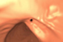
The human papilloma virus (HPV) is not associated with good health, but for some patients with advanced oropharyngeal cancer being treated with radiotherapy, the condition is beneficial, according to research presented at last week's annual meeting of the European Society for Radiotherapy and Oncology (ESTRO) in Barcelona.
Dr. Pernille Lassen.
Attendees learned that some patients with advanced oropharyngeal cancer, who are HPV-positive and are either light smokers or nonsmokers, respond to radiotherapy treatment alone without needing chemotherapy as well. Dr. Pernille Lassen, a resident in medical and radiation oncology and researcher at Aarhus University Hospital in Denmark said these patients had significantly better control of the tumor at its primary site, better disease-specific survival, and better overall survival five years after treatment than patients who were HPV negative.
Lassen and her colleagues investigated the outcomes of 181 patients on the Danish Head and Neck Cancer Group (DAHANCA) database who were treated between 1992 through 2005. They received six fractions of radiotherapy over five days together with Nimorazole, an agent that acts as a radiotherapy sensitizer to make cancer cells more receptive to the effects of radiation.
One hundred three patients had HPV-positive tumors. This group experienced better local control (81% versus 48%) than HPV-negative patients. They also had better disease-specific survival rates (90% versus 56%) and overall survival rates (77% versus 38%).
All but two of the 78 patients with HPV-negative tumors had a smoking history of more than 10 pack years, and their outcome was significantly worse than HPV positive patients, regardless of pack years. They had a disease-specific survival rate of only 50% to 52%.
Oropharyngeal cancer affects approximately 400,000 people worldwide each year. Several randomized clinical trials are now investigating treatment of patients according to HPV status.
"Although HPV status is known to be an independent prognostic factor determining tumor control and survival in radiotherapy for head and neck cancer, it is still not clear what the best treatment is for these patients. Data from the DAHANCA randomized trials has given us the opportunity to look at a group of patients, with known HPV and smoking status, who received radiotherapy without chemotherapy," explained Lassen.


![Examples of ultrasound findings and techniques. (A) Images in a 39-year-old male patient with a mass in the left thigh. The mass is heterogeneous on the B-mode US image (compared with the patient in D) and showed increased microvascularity (superb microvascular imaging [SMI]) and shear-wave elastography (SWE) values. Undifferentiated pleomorphic sarcoma was diagnosed at biopsy (with pleomorphic rhabdomyosarcoma in surgical specimen). (B) Images in an 18-year-old male patient with a mass in the left leg. The mass is hypoechoic on the B-mode image, with no other findings suggestive of malignancy. The lesion is in contact with the cortex of the tibia, which is slightly irregular. CT revealed a doubtful anteromedial tibial erosion. The microvascular study demonstrated high vascularization, suggestive of malignancy. Periosteal Ewing sarcoma was diagnosed with both histologic and immunohistochemical confirmation. (C) Images in a 69-year-old female patient with a lump growing on the outside of the left leg. Multiple SWE examinations were performed (please note the high values obtained in the measurements, whereas the color map highlights the stiffness relative to adjacent tissues). SMI showed areas of increased vascularization to target for sampling. Undifferentiated spindle cell sarcoma was diagnosed at biopsy, with residual leiomyosarcoma in the surgical specimen after neoadjuvant therapy. (D) Images in a 56-year-old female patient with a mass in the right thigh. The mass is heterogeneous at both B-mode ultrasound (similar to patient A) and MRI (coronal T2-weighted spectral attenuated inversion recovery [SPAIR]; T1-weighted pre-contrast and postcontrast imaging), which even shows uptake after the administration of paramagnetic contrast material, which is traditionally suggestive of malignancy. Low values at SMI and elastography are suggestive of benignity. Spindle cell lipoma was diagnosed at biopsy, with atypical spindle cell lipomatous tumor in the surgical specimen.](https://img.auntminnieeurope.com/mindful/smg/workspaces/default/uploads/2026/08/images-radiol250278fig2.APCFLSvX6p.jpg?auto=format%2Ccompress&fit=crop&h=100&q=70&w=100)


![Examples of ultrasound findings and techniques. (A) Images in a 39-year-old male patient with a mass in the left thigh. The mass is heterogeneous on the B-mode US image (compared with the patient in D) and showed increased microvascularity (superb microvascular imaging [SMI]) and shear-wave elastography (SWE) values. Undifferentiated pleomorphic sarcoma was diagnosed at biopsy (with pleomorphic rhabdomyosarcoma in surgical specimen). (B) Images in an 18-year-old male patient with a mass in the left leg. The mass is hypoechoic on the B-mode image, with no other findings suggestive of malignancy. The lesion is in contact with the cortex of the tibia, which is slightly irregular. CT revealed a doubtful anteromedial tibial erosion. The microvascular study demonstrated high vascularization, suggestive of malignancy. Periosteal Ewing sarcoma was diagnosed with both histologic and immunohistochemical confirmation. (C) Images in a 69-year-old female patient with a lump growing on the outside of the left leg. Multiple SWE examinations were performed (please note the high values obtained in the measurements, whereas the color map highlights the stiffness relative to adjacent tissues). SMI showed areas of increased vascularization to target for sampling. Undifferentiated spindle cell sarcoma was diagnosed at biopsy, with residual leiomyosarcoma in the surgical specimen after neoadjuvant therapy. (D) Images in a 56-year-old female patient with a mass in the right thigh. The mass is heterogeneous at both B-mode ultrasound (similar to patient A) and MRI (coronal T2-weighted spectral attenuated inversion recovery [SPAIR]; T1-weighted pre-contrast and postcontrast imaging), which even shows uptake after the administration of paramagnetic contrast material, which is traditionally suggestive of malignancy. Low values at SMI and elastography are suggestive of benignity. Spindle cell lipoma was diagnosed at biopsy, with atypical spindle cell lipomatous tumor in the surgical specimen.](https://img.auntminnieeurope.com/mindful/smg/workspaces/default/uploads/2026/08/images-radiol250278fig2.APCFLSvX6p.jpg?auto=format%2Ccompress&fit=crop&h=112&q=70&w=112)