Diffusion-weighted MRI (DWI-MRI) appears to be a very promising tool for breast cancer screening because it offers beneficial noncontrast-enhanced imaging results in a short acquisition and postprocessing time, Italian authors have found in a study published online on 13 March in European Radiology.
In addition, the group found that apparent diffusion coefficient (ADC) values obtained through DWI-MRI vary significantly according to biological tumor features.
"A deeper knowledge of how biological heterogeneity of the tumor influences imaging parameters will become critical for its clinical applications," lead study author Dr. Laura Martincich, from the Institute for Cancer Research and Treatment in Turin, and colleagues wrote.
Diffusion-weighted imaging traditionally has been used to evaluate ischemic cerebral infarction because of the MRI technique's ability to measure the mobility of water in tissue, the authors noted. This capability means there has been growing interest in DWI in cancer management.
"Currently, the role of [diffusion-weighted imaging] in the management of patients with breast diseases is still under investigation," they wrote. "However, this technique shows the potential to improve the diagnostic performance of [dynamic contrast-enhanced MRI] in the characterization of breast lesions."
 |
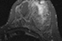
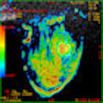
| Images are of a 45-year-old woman with HER2-enriched invasive ductal breast cancer between the external quadrants of the right breast. At diffusion-weighted MRI with a b-value of 900 sec/mm2, the cancer was identified as a hyperintense area with a cental hypointense component, suggesting intralesional necrosis (above). ADC value (1.73 x 10⁻³ mm2/sec) was obtained on an ADC map by manually tracing a region of interest within the margins of the hyperintense portion of the lesion (below). At the immunohistochemical analysis, the cancer was characterized by negative hormonal receptor status, a Ki-67 expression equal to 36%, and a HER2 status equal to 3+, classifying the HER2-enriched subtype. Images courtesy of Dr. Laura Martincich. |
 |
The prospective, single-center study enrolled 190 patients, with a mean age of 47 years (ranging from 27 to 74 years old), for local staging of their breast cancer. All the participants were recently diagnosed through biopsy with invasive breast cancer and underwent MRI examinations followed by surgery between January 2007 and December 2009.
The MRI protocol consisted of 1.5-tesla MRI scans (GE Healthcare) with both diffusion-weighted imaging and dynamic contrast-enhanced imaging (Magnevist, Bayer HealthCare Pharmaceuticals) of patients in the prone position. In addition, the MRI exams were performed at least two weeks after image-guided biopsy in order to avoid artifacts due to hemorrhage. Breast cancer patients also underwent surgery within three weeks of both types of MRI exams.
The researchers superimposed dynamic contrast-enhanced MR images onto ADC maps for cancer lesion detection and obtained ADC values by tracing a region of interest within the lesion margins.
Two pathologists with more than 15 years of experience analyzed the breast specimens. Both readers were blinded to the results of the MRI scans and defined the histological type of breast cancer based on World Health Organization criteria.
DWI results
A total of 192 breast cancers were discovered among the 190 study participants. Two patients had bilateral invasive carcinoma. Dynamic contrast-enhanced MRI identified all 192 lesions as areas of enhancement, while diffusion-weighted imaging identified all 192 lesions as hyperintense.
The mean tumor diameter was 33 mm, ranging from 6 to 100 mm. Six lesions had a contrast-enhanced MRI diameter of less than 1 cm. The mean histological tumor size was 22 mm, ranging from 6 to 65 mm.
For each of the 192 cancers, Martincich and colleagues examined the correlation between ADC and histopathological and immunohistochemical breast tumor features, such as size, type, grade, estrogen receptor status, Ki-67 expression, and HER2 status.
The images also revealed that most of the tumors (80%) were infiltrating ductal carcinomas, with histopathological grade defined in 181 (94%) of the 192 lesions. There were 60 cases (31%) of Grade 1 and Grade 2 breast cancers, and 121 patients (63%) with Grade 3 breast cancer.
In addition, the majority of tumors (79%) were estrogen receptor-positive and all the progesterone receptor-positive tumors also showed estrogen-receptor positivity. A total of 41 tumors (21%) were found to be HER2-positive by immunohistochemistry.
In the evaluation of ADC results, researchers found median ADC values to be significantly higher in estrogen receptor-negative tumors compared to estrogen receptor-positive tumors and a "weak, statistically significant" correlation between ADC values and the percentage of estrogen receptor-positive cells
Comparisons also showed ADC values were significantly higher than that of Luminal A and Luminal B/HER2-negative tumors, with a trending statistical significance with Luminal B/HER2-positive tumors.
"High ADC values were a common finding in the most biologically aggressive tumors," the authors wrote. "In fact, while ADC values were similar according to HER2 status, the subgroup of HER2-enriched tumors showed the highest ADC median value. Furthermore, the median ADC in HER2-enriched tumors was not statistically significantly different from the median value found in triple-negative tumors."
Based on the results, Martincich and colleagues concluded that diffusion-weighted MRI "appears to be a very promising tool for breast cancer screening because it does not require contrast agent administration, has both short acquisition and postprocessing time (less than 5 minutes), and provides a real quantitative functional parameter."
The researchers recommended additional studies to determine the potential future role of diffusion-weighted imaging and breast cancer management.