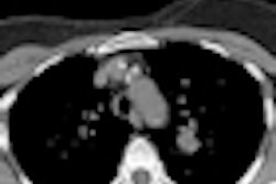
Swiss researchers have reported good results with a protocol for working up patients with nodules found on CT lung screening, according to an article in the February issue of the American Journal of Roentgenology. The algorithm produced low false-positive rates, and could make the establishment of large-scale CT screening programs more feasible.
Researchers in the International Early Lung Cancer Action Program (I-ELCAP) developed a follow-up protocol that uses shape, size, and growth rate for determining whether a lesion should be biopsied. They tested the algorithm in a study of almost 5,000 CT lung screening patients, finding that it yielded malignancies in almost three-fourths of fine-needle aspiration biopsies (FNABs), as well as 100% of bronchoscopies and one ultrasound-guided lymph nodule biopsy. Overall, less than 0.5% of CT-detected nodules were biopsied and found to be benign.
"The rate of intervention for benign nodules was indeed very low and can be further reduced," wrote Ute Wagnetz from GZO Spital Wetzikon in Switzerland, with colleagues from centers in Toronto and Freiburg, Germany (AJR, February 2012, Vol. 198:2, pp. 351-358).
Interest in CT lung screening
Lung cancer screening with CT is generating interest in more centers, particularly following last year's publication of data from the National Lung Screening Trial (NLST) showing a 20% mortality reduction among high-risk patients screened for the disease (Radiology, January 2011, Vol. 258:1, pp. 243-253).
At the same time, however, low-dose CT screening has been criticized due to the large number of false-positive results following diagnostic investigations, which could potentially increase the risks and costs of screening, diminishing the benefit of early cancer detection, Wagnetz and colleagues wrote.
The current study examined 4,782 high-risk smokers and former smokers enrolled as part of the I-ELCAP trial. Participants were required to have smoked a minimum of 10 pack years, and they had to self-report to be in good health without a history of cancer.
All participants underwent baseline low-dose CT on four- to 64-detector-row scanners from various manufacturers. Scans were performed with a low-dose technique (120 kVp and 40-60 mA) and thin-slice acquisition with 1.0- to 1.25-mm axial reconstructions. Wagnetz and colleagues reviewed the images on a PACS workstation (eFilm, Merge Healthcare) that permitted access to all prior CT studies.
The I-ELCAP diagnostic protocol defined a negative result as one in which no nodules were found that were 5 mm or larger. A positive result signified the detection of an indeterminate noncalcified solid or part-solid nodule 5 mm or larger, or a nonsolid nodule 8 mm or larger. Positive scans were followed up at defined intervals depending on nodule size and shape, with biopsy recommended immediately when noncalcified nodules reached 15 mm or larger after patients had been given antibiotics for a month with no decrease in size, per I-ELCAP recommendations.
In accordance with the I-ELCAP protocol, transthoracic fine-needle aspiration biopsy was the predominant invasive procedure in the study (n = 127), along with video-assisted thoracic surgery (VATS, n = 9). The study also saw the performance of seven bronchoscopies and one ultrasound-guided biopsy of a lymph node, as well as VATS resections for nodules not accessible for FNAB, the authors wrote. Twenty-four FNABs had unsatisfactory results, 13 of which were referred for secondary diagnostic VATS resection.
Nodule shape was most often cited as the reason to biopsy (48%), while growth on follow-up led to the decision to biopsy in 40% of the cases, and new nodules requiring biopsy led to the decision in 12% of cases. Correct biopsy indications by far outweighed incorrect ones; in all, 104 (84%) of 124 biopsies were correctly indicated and positive for malignancy, while 20 were benign (false positive), and final results were pending at publication for four cases, the authors reported.
The false-positive biopsy recommendation rate was 0.42% overall; 11.6% of FNABs and 3.6% of VATS were false positives, for an overall false-positive rate of 0.33% for FNAB and 0.10% for VATS.
Biopsy procedures recommended under I-ELCAP guidelines resulted in a low intervention rate for benign nodules, and the rate is minimal when following a research protocol that relies on shape and growth, they added, noting that the rate can be reduced further when accounting for cavitations and very fast or very slow growth rates.
"The diagnostic algorithm used in this study is based on a combined assessment of nodule shape, size, and growth," Wagnetz and colleagues wrote. "Shape as such is a rather unreliable feature for lung nodule assessment."
The authors thought it might be difficult to do a better job of finding benign nodules, owing to a lack of good criteria for their characterization. Benign nodules are associated only with calcifications and perifissural nodules, though other features have been evaluated "with limited success," Wagnetz and colleagues wrote.
"In our cohort, spiculated and lobulated solid nodules were mostly malignant; spiculations were seen only in malignant nodules," they wrote. "We did not find any other morphologic features that would have allowed us to predict the benign nature of the 20 nodules; their attenuation ranged from nonsolid to solid with smooth or lobulated margins."
Cavitations were more common in benign nodules, according to the authors. They recommended that future studies address whether the presence of cavitations can help characterize benign nodules.
The study's high rate of inconclusive FNAB results was likely due to the small size of nodules that were biopsied: nine of 10 inconclusively biopsied nodules were 10 mm or smaller, the group noted. VATS wields a greater chance of definite diagnosis for both malignancy and benign disease. For its part, thoracoscopic surgery comes with risks of bleeding, prolonged hospital stays, and chest tube duration that compare poorly with FNAB.
Any workup algorithm based on nodule shape, size, and growth will help to limit the impact of screen-detected nodules, and it might be further improved with functional imaging, Wagnetz and colleagues concluded.
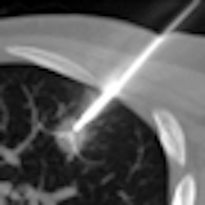
Note: Image on home page shows radiofrequency ablation of a non-small cell lung cancer with a deployable electrode needle. Image courtesy of Dr. Frédéric Deschamps from Institut Gustave Roussy in Villejuif, France.


