A trial study by German researchers has found that the use of 7-tesla MRI to image the liver can produce high-resolution, highly defined anatomical images without the need for a contrast agent.
There are both benefits and challenges for ultrahigh-field abdominal MRI, but noncontrast-enhanced angiographic applications with 7-tesla do appear promising, according to lead author Dr. Lale Umutlu, from the department of diagnostic and interventional radiology and neurology at University Hospital Essen.
While 1.5-tesla MRI has proved to be an accurate modality to image the liver, previous research also has shown the diagnostic potential of high-field MRI to image various organs and improve anatomical detail through increased sensitivity and specificity. With the introduction of 7-tesla MRI and advances in radiofrequency (RF) coil design, the authors noted that high-field MRI is expanding from neurological and musculoskeletal applications to whole-body imaging.
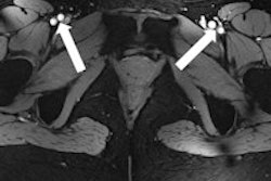
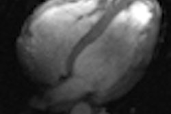
 T1-weighted 2D in (A) and opposed (B) phase MRI provides conspicuity of liver vasculature (arrows) and typical sharply defined black rims around organs with a fat/water interface in the opposed phase image (slim arrow). All images courtesy of Dr. Lale Umutlu.
Patient sample
The trial study enrolled nine healthy males and seven healthy females, with an average age of 29.5 years, ranging from 26 to 33 years of age. The 16 individuals received 7-tesla MRI examinations (Magnetom 7T, Siemens Healthcare) in a supine position.
A custom-built, eight-channel RF transmit/receive body coil also was used for image acquisition. The device consisted of two arrays with four elements placed on the upper half of the abdomen.
The researchers presented their findings in an in-press article in the European Journal of Radiology that went online last month. They acquired a series of MR images using several different techniques:
- True fast imaging with steady-state precession sequence (TrueFISP)
- T1-weighted 2D in and opposed phase sequence
- T2-weighted turbo spin-echo (TSE) sequence
- Nonenhanced T1-weighted spoiled gradient-echo sequence (2D FLASH)
- Dynamic T1-weighted spoiled gradient-echo sequence (3D FLASH) with gadobutrol (Gadovist, Bayer Schering Pharma)
- Postgadolinium T1-weighted spoiled gradient-echo sequence (2D FLASH).
The study reported no side effects from contrast-enhanced MRI exams, adding that all scans were "well tolerated." The mean MR imaging time was 37 minutes (±7 minutes), including patient positioning, shimming, and data acquisition.
Two senior radiologists with abdominal MRI experience evaluated image quality based on overall contrast resolution, sharpness, and clarity, using a five-point scale, with 1 as "insufficient/nondiagnostic" quality and 5 as "high quality."
Image evaluation
Their analysis found that of the noncontrast-enhanced MR images, 2D FLASH imaging had the highest mean scores for overall image quality (mean score of 4.52), for vessel delineation (mean score of 4.22), and for phase imaging (mean score of 4.40).
In addition, 3D FLASH was the least prone to artifacts, with a mean score of 1.33, which was significantly lower compared with TrueFISP (3.93) and TSE sequences (4.02).
Researchers also found that T2-weighted TSE imaging resulted in the greatest image impairment with a mean score of 3.92, "falling short of diagnostic relevance and precluding a clinical application." T2-weighted TSE imaging results were "significantly inferior" to both 2D FLASH and 3D FLASH images.
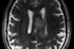
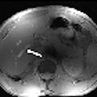
 T2-weighted TSE image is strongly impaired at 7-tesla, lacking any relevant diagnostic value.
Dynamic 3D FLASH imaging yielded good contrast enhancement of the liver and improved visualization of the liver's arteries and veins, while the portal veins were well defined in nonenhanced and all phases of dynamic imaging.
The study also found that postcontrast 2D FLASH improved conspicuity of the peripheral liver vasculature, but there was no statistically significant difference when compared to noncontrast-enhanced 2D FLASH MRI.
"Our qualitative image analysis revealed strong differences between T1-weighted and T2-weighted imaging with regards to image quality, conspicuity of anatomical details, and presence of artifacts," Umutlu and colleagues concluded. "In this study setup, 2D FLASH imaging provided best overall image quality regarding contrast resolution, sharpness, and clarity.'
In addition, the authors wrote, 7-tesla MRI created a "very good depiction of the liver vasculature with only minor improvement of conspicuity after the application of intravenous gadolinium administration."
As for future research, the authors recommended a direct comparison between 7-, 3-, and 1.5-tesla MRI to better evaluate and assess the clinical value of ultrahigh-field MRI to image the liver.