Dear AuntMinnieEurope Member,
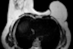
If you were to type "breast implants" into google.com, you'll get nearly 12 million search items. This underlines just how popular implants have become, not only for women seeking augmentation but also among those in need of breast reconstruction after a mastectomy.
It's important for radiologists and other medical doctors to become more familiar with the imaging of these prostheses. MRI is by far the most useful and accurate modality for these patients, according to Spanish authors who have extensive experience in this area. Click here to read more, or visit our Women's Health Digital Community.
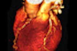
Imaging also plays a growing role in patients with acute chest pain, and the European Journal of Radiology has published a special issue on this subject. As part of the journal's coverage, researchers from the Netherlands have explained how prospective ECG-triggered cardiac CT accurately quantifies left and right ventricular function and myocardial mass. Find out more by clicking here.
Not every radiology PACS can cope with the storage and display of nuclear medicine images, and this can cause serious logistical problems. Users must check on this point before making a purchase, advises Dr. Neelam Dugar in her latest informatics column. Click here for the details.
Swiss experts in computer-assisted orthopedic surgery have developed a way of creating a patient-specific 3D model of the pelvis from a single x-ray image. This process does not require a specific calibration of the x-ray image, a computer-assisted design model of the implant, or a CT scan. Visit our Advanced Visualization Digital Community for the full story.
Visiting a hospital for an imaging examination can be a daunting experience for elderly patients, and Italian physicians think home delivery of radiology services can help overcome this problem. Click here to read about their research.
Two major congresses -- the European Society of Cardiology's annual meeting in Paris and the World Federation of Ultrasound in Medicine and Biology (WFUMB) congress in Vienna -- begin later this week. Make sure you check back starting on Friday for our coverage.


![Overview of the study design. (A) The fully automated deep learning framework was developed to estimate body composition (BC) (defined as subcutaneous adipose tissue [SAT] in liters; visceral adipose tissue [VAT] in liters; skeletal muscle [SM] in liters; SM fat fraction [SMFF] as a percentage; and intramuscular adipose tissue [IMAT] in deciliters) from MRI. The fully automated framework comprised one model (model 1) to quantify different BC measures (SAT, VAT, SM, SMFF, and IMAT) as three-dimensional (3D) measures from whole-body MRI scans. The second model (model 2) was trained to identify standardized anatomic landmarks along the craniocaudal body axis (z coordinate field), which allowed for subdividing the whole-body measures into different subregions typically examined on clinical routine MRI scans (chest, abdomen, and pelvis). (B) BC was quantified from whole-body MRI in over 66,000 individuals from two large population-based cohort studies, the UK Biobank (UKB) (36,317 individuals) and the German National Cohort (NAKO) (30,291 individuals). Bar graphs show age distribution by sex and cohort. BMI = body mass index. (C) After the performance assessment of the fully automated framework, the change in BC measures, distributions, and profiles across age decades were investigated. Age-, sex-, and height-adjusted body composition reference curves were calculated and made publicly available in a web-based z-score calculator (https://circ-ml.github.io).](https://img.auntminnieeurope.com/mindful/smg/workspaces/default/uploads/2026/05/body-comp.XgAjTfPj1W.jpg?auto=format%2Ccompress&dpr=2&fit=crop&h=167&q=70&w=250)


