A certain type of prospective ECG-triggered cardiac CT accurately quantifies left and right ventricular function and myocardial mass in comparison with cardiac MRI, researchers of an online study in the European Journal of Radiology find. In addition, the method results in substantially lower radiation exposure than traditional retrospective ECG-gating.
As part of a special EJR issue focusing on imaging the chest, European and U.S. researchers collaborated on a study to prospectively evaluate the accuracy of left and right ventricular function and myocardial mass measurements based on a dual-step, low-radiation dose protocol with prospectively ECG-triggered second generation dual-source CT using cardiac MRI as the reference standard (EJR, article in press, 9 August 2011).
Accurate determination of ventricular size, mass, and function is important for clinical diagnosis and risk assessment in the care of patients with various pathological conditions, wrote Dr. Richard Takx, from the department of radiology at Maastricht University Medical Centre in the Netherlands, and colleagues. Examples include classically left-sided heart diseases, such as coronary artery disease or left ventricular hypertrophy, which has been demonstrated as an independent risk factor for adverse cardiovascular events and premature death, added Takx, who is also affiliated with the department of radiology and radiological science at the Medical University of South Carolina in Charleston.
Assessing right ventricular function and myocardial mass aids in the evaluation of patients with a variety of right-sided heart diseases, such as pulmonary artery hypertension and chronic thromboembolic pulmonary hypertension, the authors wrote.
"In recent years, considerable technical developments of different imaging modalities have significantly improved their performance in the evaluation of ventricular mass and function," they stated.
Cardiac MRI is considered the reference standard due to its unmatched accuracy and reproducibility in the evaluation of left and right ventricle function and myocardial mass. However, in patients with contraindications for cardiac MRI, alternative imaging modalities such as echocardiography and CT must be considered. Most protocols currently use CT with retrospective ECG-gating, which can result in a substantial radiation exposure of about 14.8 to 21.1 mSv, they added. The current study sought to assess how well second-generation dual-source CT with a low radiation dose, prospectively ECG-triggered dual-step pulsing protocol measured up.
The team's study included 20 patients, who all underwent 1.5-tesla MRI (Magnetom Avanto, Siemens Healthcare) and prospectively ECG-triggered dual-step pulsing cardiac dual-source CT (Somatom Definition Flash, Siemens Healthcare).
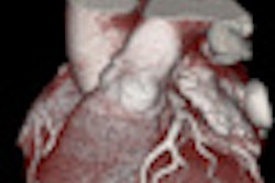
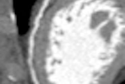
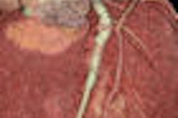
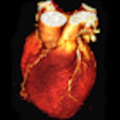
 Prospectively ECG-triggered coronary CT angiography of a 61-year-old man with chest pain. 3D-volume-rendered reconstruction shows heavily calcified coronary arteries (A). Multiplanar reformations of the left ascending coronary artery (B and C) and the right coronary artery (D) revealed multiple coronary artery stenoses of greater than 50%. Curved multiplanar reconstruction of the circumflex artery shows a soft and mixed plaque leading to a luminal narrowing of approximately 50%. All images courtesy of of Drs. Christian Fink and Thomas Henzler.
The image acquisition mode performed low-radiation (20% tube current) imaging over the majority of the cardiac cycle and applied full radiation only during a single adjustable phase. Full-radiation-phase images were used to assess cardiac morphology, while low-radiation-phase images were used to measure left and right ventricular function and mass.
The researchers compared quantitative CT measurements based on contiguous multiphase short-axis reconstructions from the axial CT data with short-axis steady-state free precession cardiac cine MRI. They also manually traced contours around the ventricular borders for calculation of left and right ventricular end-diastolic volume, end-systolic volume, stroke volume, ejection fraction, and myocardial mass for both modalities.
One experienced radiologist, with five years of clinical experience in cardiac radiology, evaluated the dual-source CT and cardiac MRI datasets and determined end-diastolic volume, end-systolic volume, stroke volume, ejection fraction, and myocardial mass for the left and right ventricles using Argus (Syngo VE36A, Wizard, Siemens Healthcare) software according to Simpson's rule.
What the researchers found is all CT measurements of left and right ventricular function and mass correlated well with those from MRI. For left/right end-diastolic volume r = 0.885/0.801, left/right end-systolic volume r = 0.947/0.879, left/right stroke volume r = 0.620/0.697, left/right ejection fraction r = 0.869/0.751, and left/right myocardial mass r = 0.959/0.702. The mean radiation dose was 6.2 ± 1.8 mSv.
"Prospective ECG-triggered scan protocols are an effective technique to markedly reduce radiation dose in cardiac CT," the authors wrote. Especially since dose for retrospective spiral ECG gating ranges between 14.8 and 21.1 mSv.
They also noted there was excellent correlation between CT and MRI for left ventricle end-diastolic volume, end-systolic volume, ejection fraction, and myocardial mass. There was good correlation for left ventricle systolic volume. For right ventricle measurements, there was excellent correlation between CT and MRI for end-diastolic volume and end-systolic volume. There was good correlation for systolic volume, ejection fraction, and myocardial mass. The good correlation may be explained by the fact the patients did not receive beta-blockers, according to the researchers.
They are also quick to point out there are several limitations to their study -- the size of the population and also pathological correlation. In addition, all patients had a normal sinus rhythm so they were unable to assess the performance of the segmentation protocol and the resulting radiation dose in patients with arrhythmia.
It should be noted one of the authors has a conflict of interest because he acts as a consultant for and receives research support from Bayer-Schering, Bracco, General Electric, Medrad, and Siemens. None of the other authors, however, have anything to disclose.