Dear AuntMinnieEurope Member,
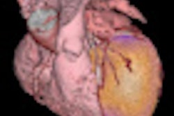
Research groups in Europe and North America are breaking new boundaries by investigating the feasibility of 7-tesla MRI for regions outside of the brain, particularly cardiac imaging. These efforts are prompted by the unmet clinical needs and unsolved technical problems of today's clinical cardiac MRI.
We asked guest columnist Dr. Thoralf Niendorf from the renowned Charité Hospital in Berlin, to investigate whether ultrafast cardiac MR represents hype or hope. His views are challenging and controversial. To find out more, click here.
Virtual surgery and 3D modeling also has cardiac applications, and a rising star in the field, Dr. Andrew Taylor from London, gave a keynote lecture on this topic at the recent British Institute of Radiology (BIR) President's Conference. Click here for more details.
For additional stories on the fast-moving area of cardiac imaging, take a look at our new Cardiac Imaging Digital Community, which launched earlier this week. We promise to bring you a diverse selection of other articles during the coming weeks and months.
Early results from the third U.K. CT dose survey caused quite a stir this week at the U.K. Radiological Congress (UKRC) in Manchester. This is the first national survey conducted since 2003, so it has been eagerly awaited, and an electronic poster on this subject was the most popular exhibit during the first two days of the congress. For the full story, click here.
After the terrible recent E. coli outbreak in Germany, salads were off the menu at the congress venue for the Deutscher Röntgenkongress (DRK) in Hamburg, but there was still plenty of informative and educational material to offer. Dr. Matthias Dietzel reveals all in his congress report, and you can also read about the experiences of two first-time attendees by clicking here.
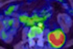
Across the Atlantic, speakers from across the globe have been presenting their research at the Society of Nuclear Medicine (SNM)'s annual meeting. For the inside track on the hot clinical topics, click here.


![Overview of the study design. (A) The fully automated deep learning framework was developed to estimate body composition (BC) (defined as subcutaneous adipose tissue [SAT] in liters; visceral adipose tissue [VAT] in liters; skeletal muscle [SM] in liters; SM fat fraction [SMFF] as a percentage; and intramuscular adipose tissue [IMAT] in deciliters) from MRI. The fully automated framework comprised one model (model 1) to quantify different BC measures (SAT, VAT, SM, SMFF, and IMAT) as three-dimensional (3D) measures from whole-body MRI scans. The second model (model 2) was trained to identify standardized anatomic landmarks along the craniocaudal body axis (z coordinate field), which allowed for subdividing the whole-body measures into different subregions typically examined on clinical routine MRI scans (chest, abdomen, and pelvis). (B) BC was quantified from whole-body MRI in over 66,000 individuals from two large population-based cohort studies, the UK Biobank (UKB) (36,317 individuals) and the German National Cohort (NAKO) (30,291 individuals). Bar graphs show age distribution by sex and cohort. BMI = body mass index. (C) After the performance assessment of the fully automated framework, the change in BC measures, distributions, and profiles across age decades were investigated. Age-, sex-, and height-adjusted body composition reference curves were calculated and made publicly available in a web-based z-score calculator (https://circ-ml.github.io).](https://img.auntminnieeurope.com/mindful/smg/workspaces/default/uploads/2026/05/body-comp.XgAjTfPj1W.jpg?auto=format%2Ccompress&dpr=2&fit=crop&h=167&q=70&w=250)


