Dear MRI Insider,
Welcome to the first edition of the AuntMinnieEurope.com MRI Insider. This email newsletter will provide regular updates on the latest MRI developments in Europe, as reported in our new MRI Digital Community.
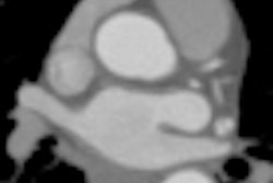
It's certainly been a hectic month for MRI, in terms of both congress presentations and original research articles. At the British Institute of Radiology (BIR) President's Conference in London, consultant cardiologist Dr. Mark Westwood explained how cardiac MR can produce fast, clinically useful results, particularly with cooperative patients. Technique is all-important, he noted. For the full story, click here.
Radiologists from Jena, Germany, have found that the fusion of 3-tesla MR images with x-ray mammograms can lead to clinically significant improvements in clinical workflow and diagnostic accuracy. They have shown that the semiautomatic image registration of x-ray mammography and high-field MRI is both feasible and beneficial. Click here for the details.
European researchers were active participants at last month's International Society for Magnetic Resonance in Medicine (ISMRM) meeting in Montreal, Canada. For instance, a group from the University of Bonn, Germany, revealed that MR angiography with blood-pool contrast can help simultaneously detect and assess arterial and additional venous conditions. Click here to read staff writer Wayne Forrest's onsite report.
New guidelines from the European Society of Urogenital Radiology on dealing with uterine cervical cancer patients have confirmed the value of MRI as the imaging modality of choice for preoperative staging and follow-up. The lead author of the guidelines is Dr. Corinne Balleyguier from Paris, who is a member of AuntMinnieEurope.com's editorial advisory board. To read more, click here.
Tackling the medical issue of unexplained nipple discharge can prove especially challenging. Italian researchers emphasize that breast MRI can find additional cancers, not recognized or occult when conventional imaging is used. Click here to find out more.
Finally, make sure you check out the MRI Digital Community next week for a guest column about how ultrafast MR systems can be used successfully in cardiac patients.


![Examples of ultrasound findings and techniques. (A) Images in a 39-year-old male patient with a mass in the left thigh. The mass is heterogeneous on the B-mode US image (compared with the patient in D) and showed increased microvascularity (superb microvascular imaging [SMI]) and shear-wave elastography (SWE) values. Undifferentiated pleomorphic sarcoma was diagnosed at biopsy (with pleomorphic rhabdomyosarcoma in surgical specimen). (B) Images in an 18-year-old male patient with a mass in the left leg. The mass is hypoechoic on the B-mode image, with no other findings suggestive of malignancy. The lesion is in contact with the cortex of the tibia, which is slightly irregular. CT revealed a doubtful anteromedial tibial erosion. The microvascular study demonstrated high vascularization, suggestive of malignancy. Periosteal Ewing sarcoma was diagnosed with both histologic and immunohistochemical confirmation. (C) Images in a 69-year-old female patient with a lump growing on the outside of the left leg. Multiple SWE examinations were performed (please note the high values obtained in the measurements, whereas the color map highlights the stiffness relative to adjacent tissues). SMI showed areas of increased vascularization to target for sampling. Undifferentiated spindle cell sarcoma was diagnosed at biopsy, with residual leiomyosarcoma in the surgical specimen after neoadjuvant therapy. (D) Images in a 56-year-old female patient with a mass in the right thigh. The mass is heterogeneous at both B-mode ultrasound (similar to patient A) and MRI (coronal T2-weighted spectral attenuated inversion recovery [SPAIR]; T1-weighted pre-contrast and postcontrast imaging), which even shows uptake after the administration of paramagnetic contrast material, which is traditionally suggestive of malignancy. Low values at SMI and elastography are suggestive of benignity. Spindle cell lipoma was diagnosed at biopsy, with atypical spindle cell lipomatous tumor in the surgical specimen.](https://img.auntminnieeurope.com/mindful/smg/workspaces/default/uploads/2026/08/images-radiol250278fig2.APCFLSvX6p.jpg?auto=format%2Ccompress&fit=crop&h=100&q=70&w=100)


![Examples of ultrasound findings and techniques. (A) Images in a 39-year-old male patient with a mass in the left thigh. The mass is heterogeneous on the B-mode US image (compared with the patient in D) and showed increased microvascularity (superb microvascular imaging [SMI]) and shear-wave elastography (SWE) values. Undifferentiated pleomorphic sarcoma was diagnosed at biopsy (with pleomorphic rhabdomyosarcoma in surgical specimen). (B) Images in an 18-year-old male patient with a mass in the left leg. The mass is hypoechoic on the B-mode image, with no other findings suggestive of malignancy. The lesion is in contact with the cortex of the tibia, which is slightly irregular. CT revealed a doubtful anteromedial tibial erosion. The microvascular study demonstrated high vascularization, suggestive of malignancy. Periosteal Ewing sarcoma was diagnosed with both histologic and immunohistochemical confirmation. (C) Images in a 69-year-old female patient with a lump growing on the outside of the left leg. Multiple SWE examinations were performed (please note the high values obtained in the measurements, whereas the color map highlights the stiffness relative to adjacent tissues). SMI showed areas of increased vascularization to target for sampling. Undifferentiated spindle cell sarcoma was diagnosed at biopsy, with residual leiomyosarcoma in the surgical specimen after neoadjuvant therapy. (D) Images in a 56-year-old female patient with a mass in the right thigh. The mass is heterogeneous at both B-mode ultrasound (similar to patient A) and MRI (coronal T2-weighted spectral attenuated inversion recovery [SPAIR]; T1-weighted pre-contrast and postcontrast imaging), which even shows uptake after the administration of paramagnetic contrast material, which is traditionally suggestive of malignancy. Low values at SMI and elastography are suggestive of benignity. Spindle cell lipoma was diagnosed at biopsy, with atypical spindle cell lipomatous tumor in the surgical specimen.](https://img.auntminnieeurope.com/mindful/smg/workspaces/default/uploads/2026/08/images-radiol250278fig2.APCFLSvX6p.jpg?auto=format%2Ccompress&fit=crop&h=112&q=70&w=112)