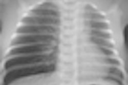
Researchers are advising healthcare providers to take extra care in the use of x-ray procedures in pregnant women and very young children, following results of a study published online February 10 in BMJ of more than 7,500 U.K. children who received radiography procedures.
The researchers found a slightly higher risk of developing cancer after x-ray exams, although the data were not statistically significant for any cancer except lymphoma. Still, the researchers took a "better safe than sorry" stance in advising caution.
The authors said they wanted to follow up research from other studies, including a 1997 study on the Oxford Survey of Childhood Cancers, which found a 40% increase in cancer in children whose mothers were exposed to abdominal radiography while pregnant during the years 1953 to 1981.
The new study, the U.K. Childhood Cancer Study, analyzed the medical records of 2,690 children who developed cancer and 4,858 healthy children matched by age, sex, and region, all of whom were born in England and Wales over a 30-year time span from 1976 to 1996 (BMJ, February 10, 2011).
A total of 305 children were exposed to 319 examinations that emitted radiation dose while in utero, and 170 children received 247 diagnostic x-ray examinations in early infancy. Researchers from the U.S. National Cancer Institute's Division of Cancer Epidemiology and Genetics in Bethesda, MD, and the University of York in York, U.K., measured the risk of developing leukemia, lymphoma, and central nervous system tumors, as well as the risk of developing childhood cancer overall.
Lead author Preetha Rajaraman, PhD, and colleagues reported that in utero exposure produced a slightly higher risk of developing any cancer (1.14 odds ratio), with the greatest risk of developing leukemia (1.36). These risk levels were not statistically significant, however.
Exposure to diagnostic x-rays when younger than 3 months of age was associated with a small, nonsignificant extra risk for all cancers (1.16 odds ratio) and leukemia (1.63). The risk of developing lymphoma was five times greater and was statistically significant, but this finding was based on only seven cases, and these children were either low birth weight or were born prematurely.
The authors noted that the risk levels found in their study were lower than those of the Oxford study, which could be due an ongoing trend of lower radiation dose per image and fewer images per patient. The prevalence of radiography in the current study in the 1980s and 1990s was 3.8%, compared with 10% to 15% in the Oxford survey in the 1950s to 1980s.
The researchers added that their data showed no association between exposure to ultrasound scans and risk of childhood cancer. The lack of risk for ultrasound argues against a systematic bias in the study, the authors said, and suggests that the current x-ray risk statistics, while not statistically significant, "could indicate a picture of modestly elevated risk" for radiography.
By Cynthia E. Keen
AuntMinnie.com staff writer
February 11, 2011


![Examples of ultrasound findings and techniques. (A) Images in a 39-year-old male patient with a mass in the left thigh. The mass is heterogeneous on the B-mode US image (compared with the patient in D) and showed increased microvascularity (superb microvascular imaging [SMI]) and shear-wave elastography (SWE) values. Undifferentiated pleomorphic sarcoma was diagnosed at biopsy (with pleomorphic rhabdomyosarcoma in surgical specimen). (B) Images in an 18-year-old male patient with a mass in the left leg. The mass is hypoechoic on the B-mode image, with no other findings suggestive of malignancy. The lesion is in contact with the cortex of the tibia, which is slightly irregular. CT revealed a doubtful anteromedial tibial erosion. The microvascular study demonstrated high vascularization, suggestive of malignancy. Periosteal Ewing sarcoma was diagnosed with both histologic and immunohistochemical confirmation. (C) Images in a 69-year-old female patient with a lump growing on the outside of the left leg. Multiple SWE examinations were performed (please note the high values obtained in the measurements, whereas the color map highlights the stiffness relative to adjacent tissues). SMI showed areas of increased vascularization to target for sampling. Undifferentiated spindle cell sarcoma was diagnosed at biopsy, with residual leiomyosarcoma in the surgical specimen after neoadjuvant therapy. (D) Images in a 56-year-old female patient with a mass in the right thigh. The mass is heterogeneous at both B-mode ultrasound (similar to patient A) and MRI (coronal T2-weighted spectral attenuated inversion recovery [SPAIR]; T1-weighted pre-contrast and postcontrast imaging), which even shows uptake after the administration of paramagnetic contrast material, which is traditionally suggestive of malignancy. Low values at SMI and elastography are suggestive of benignity. Spindle cell lipoma was diagnosed at biopsy, with atypical spindle cell lipomatous tumor in the surgical specimen.](https://img.auntminnieeurope.com/mindful/smg/workspaces/default/uploads/2026/08/images-radiol250278fig2.APCFLSvX6p.jpg?auto=format%2Ccompress&fit=crop&h=100&q=70&w=100)


![Examples of ultrasound findings and techniques. (A) Images in a 39-year-old male patient with a mass in the left thigh. The mass is heterogeneous on the B-mode US image (compared with the patient in D) and showed increased microvascularity (superb microvascular imaging [SMI]) and shear-wave elastography (SWE) values. Undifferentiated pleomorphic sarcoma was diagnosed at biopsy (with pleomorphic rhabdomyosarcoma in surgical specimen). (B) Images in an 18-year-old male patient with a mass in the left leg. The mass is hypoechoic on the B-mode image, with no other findings suggestive of malignancy. The lesion is in contact with the cortex of the tibia, which is slightly irregular. CT revealed a doubtful anteromedial tibial erosion. The microvascular study demonstrated high vascularization, suggestive of malignancy. Periosteal Ewing sarcoma was diagnosed with both histologic and immunohistochemical confirmation. (C) Images in a 69-year-old female patient with a lump growing on the outside of the left leg. Multiple SWE examinations were performed (please note the high values obtained in the measurements, whereas the color map highlights the stiffness relative to adjacent tissues). SMI showed areas of increased vascularization to target for sampling. Undifferentiated spindle cell sarcoma was diagnosed at biopsy, with residual leiomyosarcoma in the surgical specimen after neoadjuvant therapy. (D) Images in a 56-year-old female patient with a mass in the right thigh. The mass is heterogeneous at both B-mode ultrasound (similar to patient A) and MRI (coronal T2-weighted spectral attenuated inversion recovery [SPAIR]; T1-weighted pre-contrast and postcontrast imaging), which even shows uptake after the administration of paramagnetic contrast material, which is traditionally suggestive of malignancy. Low values at SMI and elastography are suggestive of benignity. Spindle cell lipoma was diagnosed at biopsy, with atypical spindle cell lipomatous tumor in the surgical specimen.](https://img.auntminnieeurope.com/mindful/smg/workspaces/default/uploads/2026/08/images-radiol250278fig2.APCFLSvX6p.jpg?auto=format%2Ccompress&fit=crop&h=112&q=70&w=112)