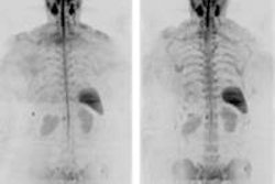
NEW YORK (Reuters Health), Mar 18 - By detecting focal lesions in bone, whole-body MRI can help predict the risk of progression from asymptomatic to symptomatic multiple myeloma (MM), German researchers report in the Journal of Clinical Oncology published online on February 22.
They say that the presence, and especially the number, of focal lesions were the strongest predictors of progression to symptomatic disease. Their study builds on earlier work showing that axial MRI (spine and sacrum only) has prognostic value in symptomatic MM.
For the first five years, the progression rate from asymptomatic to symptomatic MM is 10% per year, and by 15 years roughly three-quarters of patients have progressed to symptomatic disease, according to the researchers.
The senior author, Dr. Hartmut Goldschmidt of University Hospital Heidelberg, and his colleagues recommend whole-body MRI to stratify patients' risk. Once identified, patients with focal lesions or other high-risk features could be included in clinical trials testing therapies such as lenalidomide, Dr. Goldschmidt told Reuters Health by e-mail.
On the other hand, the paper cautions that prospective studies are needed to determine "whether patients in whom MRI shows lesions will benefit from a treatment that they would not receive if only standard imaging protocols were used."
The study included 149 patients (median age 58) with asymptomatic MM, none of whom received systemic treatment until symptoms developed. The median follow-up was 23.7 months.
The authors discovered focal lesions in 42 patients (28%). The number of lesions per patient ranged from one to more than 20. Patients with more than one focal lesion had significantly shorter progression-free survival (median, 13 months) compared to patients with one or no focal lesion (median not reached, with last event at 43 months; p < 0.001).
On multivariate analysis, the presence and number of focal lesions and the presence of diffuse bone marrow infiltration were the only significant predictors of progression to symptomatic disease.
In 30 patients, lesions outside the axial area would have been missed on axial MRI, the authors point out. In 19 patients, all of the lesions were outside the axial area.
The report notes that properly equipped, modern MRI scanners can scan the whole body in 30 to 45 minutes.
By Scott Baltic
J Clin Oncol 2010.
Last Updated: 2010-03-17 19:43:50 -0400 (Reuters Health)
Related Reading
Contrast MRI helps guide stem cell therapy for myeloma, February 11, 2010
New progress in multiple myeloma treatment; MDCT has edge in detection, June 18, 2007
High-dose therapy does not improve multiple myeloma outcome, March 9, 2006
No survival benefit of high-dose therapy in multiple myeloma, January 5, 2006
Copyright © 2010 Reuters Limited. All rights reserved. Republication or redistribution of Reuters content, including by framing or similar means, is expressly prohibited without the prior written consent of Reuters. Reuters shall not be liable for any errors or delays in the content, or for any actions taken in reliance thereon. Reuters and the Reuters sphere logo are registered trademarks and trademarks of the Reuters group of companies around the world.