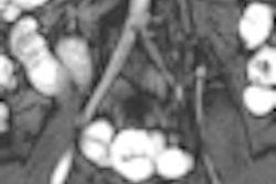
Although MRI enterography can diagnose important incidental findings in a minority of patients with suspected or known Crohn's disease, Dutch researchers have concluded that a substantial number of patients experience unnecessary morbidity because of additional examinations of benign or normal conditions.
In the study, published in the January issue of the World Journal of Gastroenterology, MRI enterography revealed incidental findings outside the small intestine that were not related to Crohn's disease in 25% of patients. As a result, additional examinations were given to 5% of patients (2010, Vol. 16:1, pp. 76-82).
"The detection rate of important incidental lesions not related to Crohn's disease was too low to be an argument in itself for performing MRI enterography in this group of patients," wrote lead author Dr. Michael Dam Jensen from the department of internal medicine at Vejle Hospital in Vejle, Denmark, and colleagues.
MRI has been used increasingly to assess small-bowel Crohn's disease, because the modality helps view the disease beyond the intestinal wall. MRI also can find some unexpected extraintestinal findings that are unrelated to Crohn's disease.
Increased morbidity
"On one hand," the authors noted, "modern imaging techniques may detect early extraintestinal malignant disease or disease requiring clinical intervention, thereby reducing morbidity and mortality. On the other hand, incidental findings may lead to further diagnostic workup or surgery of benign lesions causing increased morbidity."
The retrospective study collected data from December 2003 to November 2007 of patients with suspected or known Crohn's disease, symptoms consistent with disease activity, or related complications.
All patients received MRI enterography. Scans were conducted in the supine position on a 1.5-tesla MRI system (Intera, Philips Healthcare, Andover, MA) with a body coil (Philips). Gadodiamide (0.1 mmol/kg) also was administered (Omniscan, GE Healthcare, Chalfont St. Giles, U.K.) intravenously to the patients.
The researchers evaluated unexpected findings outside the small intestine not previously known or suspected at the time of referral and unrelated to inflammatory bowel disease.
The study excluded 29 scans that were performed for indications other than inflammatory bowel disease, two scans from patients who declined to take a contrast agent, and 40 scans from 29 patients because of repeated MRI enterographies in the study period.
Patient population
The final analysis included 283 MRI enterography exams from 283 patients. There were 193 females (68%) among the 283 patients, who had a mean age of 38.7 years, ranging from 10 to 85 years.
MRI suspected Crohn's disease in 156 patients (55%), with newly diagnosed or known Crohn's disease in 127 cases (45%). The modality also discovered active Crohn's disease in 88 patients (31%), fistula in four patients (1.4%), and abscess in two patients (0.7%). There was no difference in mean age between patients with known and suspected Crohn's disease.
Extraintestinal findings unrelated to Crohn's disease were recorded in 72 patients (25%), of which 58 patients (20%) had 74 previously unknown lesions. Important or incompletely characterized findings were detected in 17 patients (6%).
Incidental findings led to 12 further interventions in nine patients (3.2%), revealing previously unknown pathological conditions in five cases (1.8%). One patient underwent surgery and one patient was diagnosed with a malignant disease.
Unnecessary examinations
MRI detected incidental colonic lesions in 16 patients, of which additional workup in four cases revealed normal anatomy. Two patients benefited from the additional examinations, but incidental findings led to unnecessary examinations in nine patients (3.2%).
The group concluded that while incidental findings were common in patients with known and suspected Crohn's disease with MRI, additional examinations revealed important disease "in only a minority of patients. However, a substantial number of patients experienced unnecessary morbidity because of the additional examinations of benign or normal conditions. The detection rate of important incidental lesions not related to [Crohn's disease] was too low to be an argument in itself for performing MRI enterography in this group of patients."
The study was limited by its retrospective approach, according to the researchers, as "radiological reports were not performed with the focus on incidental findings, and underestimation of clinically unimportant findings are likely."
The inclusion of more women than men in the study may have been a limitation, though the authors added that Crohn's disease is more common in females. They also cited previous research that the prevalence of women with irritable bowel syndrome is as much as four times greater than men.
By Wayne Forrest
AuntMinnie.com staff writer
February 23, 2010
Related Reading
ASIR cuts dose in Crohn's disease patients, December 4, 2009
FDG-PET/CT helps manage treatment for Crohn's disease, November 16, 2009
Colonoscopy findings usually not needed for Crohn's treatment decisions, September 28, 2009
Musculoskeletal deficits associated with pediatric Crohn's, January 29, 2009
Crohn's disease patients at risk of significant diagnostic radiation exposure, November 18, 2008
Copyright © 2010 AuntMinnie.com