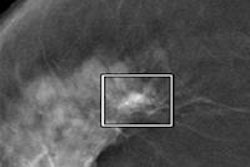
Full-field digital mammography (FFDM) can facilitate increased detection of smaller tumors compared to conventional mammography. But adding computer-aided detection (CAD) technology to FFDM can yield even better results, according to researchers at the Hospital Virgen de la Salud in Toledo, Spain.
The Spanish study team also found that while the overall increase of detected cancers from FFDM and CAD were not statistically significant, CAD did lead to a significant increase in the proportion of in situ cancers. In addition, CAD increased the number of biopsies, owing to its ability to detect more microcalcifications, said Dr. Mabel Garcia-Hidalgo.
Garcia-Hidalgo presented the research on behalf of lead author Dr. Cristina Romero at the 2009 RSNA meeting in Chicago.
The researchers sought to assess the impact of FFDM and CAD on their institution's multidisciplinary breast unit. A retrospective study was performed to evaluate three different modes of reading: conventional mammography, FFDM alone, and the addition of CAD to all FFDM exams.
The conventional mammography approach was evaluated over a seven-month period in 2004, while FFDM and FFDM + CAD were evaluated over seven-month periods in 2006 and 2007, respectively.
All three phases included both screening and diagnostic mammograms. In the study period in 2004, 73.8% of the 4,064 mammograms were screening studies, while 74.4% of the 4,205 mammograms during the FFDM period were screening exams. In the FFDM + CAD period in 2007, 67.6% of the 5,184 were screening studies.
The women all received a standard two-projection baseline mammogram, and there were no significant differences in patient ages during the three study periods.
In cancer cases, the researchers only included studies with visible findings on mammograms. Patients with BI-RADS 4 and 5 ratings received biopsies, and MRI was performed in all breast cancer patients, according to the researchers.
FFDM and FFDM + CAD both enabled the detection of smaller tumors at diagnosis.
Number of tumors found per modality, by tumor size
|
The researchers also found that FFDM + CAD produced a significant difference in the proportion of carcinomas detected in situ compared to conventional mammography (p = 0.05). Of the detected cancers, 17.8% were in situ with FFDM + CAD, while 10.3% were in situ during the conventional mammography period.
In the FFDM-only period, 14.6% of detected cancers were in situ. The difference was not statistically significant, however.
In the FFDM + CAD period, 11.2% of patients received biopsies, compared with 8.15% with FFDM only and 10.21% with conventional mammography. The differences were statistically significant (p = 0.02).
Total cancer detection increased from 39 (9.3%) with conventional mammography to 48 with FFDM (11.4%) and 74 (16.9%). However, the differences were not statistically significant (p = 0.07).
In other results, the researchers found no statistical differences in the distribution of radiological findings between the three techniques.
By Erik L. Ridley
AuntMinnie.com staff writer
February 26, 2010
Related Reading
Ignoring breast CAD detections cuts sensitivity, October 23, 2009
Breast ultrasound CAD helps doctors find smaller lesions, October 10, 2008
CARS report: CAD prompts range of behaviors in readers, June 26, 2008
Breast CAD improves classification accuracy, cuts ultrasound biopsies, February 29, 2008
Breast MRI CAD doesn't improve accuracy due to poor DCIS detection, February 18, 2008
Copyright © 2010 AuntMinnie.com


![Examples of ultrasound findings and techniques. (A) Images in a 39-year-old male patient with a mass in the left thigh. The mass is heterogeneous on the B-mode US image (compared with the patient in D) and showed increased microvascularity (superb microvascular imaging [SMI]) and shear-wave elastography (SWE) values. Undifferentiated pleomorphic sarcoma was diagnosed at biopsy (with pleomorphic rhabdomyosarcoma in surgical specimen). (B) Images in an 18-year-old male patient with a mass in the left leg. The mass is hypoechoic on the B-mode image, with no other findings suggestive of malignancy. The lesion is in contact with the cortex of the tibia, which is slightly irregular. CT revealed a doubtful anteromedial tibial erosion. The microvascular study demonstrated high vascularization, suggestive of malignancy. Periosteal Ewing sarcoma was diagnosed with both histologic and immunohistochemical confirmation. (C) Images in a 69-year-old female patient with a lump growing on the outside of the left leg. Multiple SWE examinations were performed (please note the high values obtained in the measurements, whereas the color map highlights the stiffness relative to adjacent tissues). SMI showed areas of increased vascularization to target for sampling. Undifferentiated spindle cell sarcoma was diagnosed at biopsy, with residual leiomyosarcoma in the surgical specimen after neoadjuvant therapy. (D) Images in a 56-year-old female patient with a mass in the right thigh. The mass is heterogeneous at both B-mode ultrasound (similar to patient A) and MRI (coronal T2-weighted spectral attenuated inversion recovery [SPAIR]; T1-weighted pre-contrast and postcontrast imaging), which even shows uptake after the administration of paramagnetic contrast material, which is traditionally suggestive of malignancy. Low values at SMI and elastography are suggestive of benignity. Spindle cell lipoma was diagnosed at biopsy, with atypical spindle cell lipomatous tumor in the surgical specimen.](https://img.auntminnieeurope.com/mindful/smg/workspaces/default/uploads/2026/08/images-radiol250278fig2.APCFLSvX6p.jpg?auto=format%2Ccompress&fit=crop&h=100&q=70&w=100)


![Examples of ultrasound findings and techniques. (A) Images in a 39-year-old male patient with a mass in the left thigh. The mass is heterogeneous on the B-mode US image (compared with the patient in D) and showed increased microvascularity (superb microvascular imaging [SMI]) and shear-wave elastography (SWE) values. Undifferentiated pleomorphic sarcoma was diagnosed at biopsy (with pleomorphic rhabdomyosarcoma in surgical specimen). (B) Images in an 18-year-old male patient with a mass in the left leg. The mass is hypoechoic on the B-mode image, with no other findings suggestive of malignancy. The lesion is in contact with the cortex of the tibia, which is slightly irregular. CT revealed a doubtful anteromedial tibial erosion. The microvascular study demonstrated high vascularization, suggestive of malignancy. Periosteal Ewing sarcoma was diagnosed with both histologic and immunohistochemical confirmation. (C) Images in a 69-year-old female patient with a lump growing on the outside of the left leg. Multiple SWE examinations were performed (please note the high values obtained in the measurements, whereas the color map highlights the stiffness relative to adjacent tissues). SMI showed areas of increased vascularization to target for sampling. Undifferentiated spindle cell sarcoma was diagnosed at biopsy, with residual leiomyosarcoma in the surgical specimen after neoadjuvant therapy. (D) Images in a 56-year-old female patient with a mass in the right thigh. The mass is heterogeneous at both B-mode ultrasound (similar to patient A) and MRI (coronal T2-weighted spectral attenuated inversion recovery [SPAIR]; T1-weighted pre-contrast and postcontrast imaging), which even shows uptake after the administration of paramagnetic contrast material, which is traditionally suggestive of malignancy. Low values at SMI and elastography are suggestive of benignity. Spindle cell lipoma was diagnosed at biopsy, with atypical spindle cell lipomatous tumor in the surgical specimen.](https://img.auntminnieeurope.com/mindful/smg/workspaces/default/uploads/2026/08/images-radiol250278fig2.APCFLSvX6p.jpg?auto=format%2Ccompress&fit=crop&h=112&q=70&w=112)