Virtual colonoscopy computer-aided detection (CAD) can improve sensitivity for polyp detection, especially for the inexperienced reader, according to a study from Paris.
The team, from Hôpital Pitié-Salpêtrière-Assistance Publique-Hôpitaux de Paris, sought to determine if polyp detection could be improved with the aid of an investigational CAD application.
The study included 40 subjects (14 men and 26 women, ages 45-76, mean 62) who all had an above-average risk of colorectal polyps and cancers, including 42% with iron-deficiency anemia, 23% with chronic abdominal pain, and 35% with a family history of colorectal cancer.
All patients followed a liquid diet and cathartic bowel preparation (Phospho-soda, Fleet Pharmaceuticals, Lynchburg, VA; and two 45-mL bisacodyl tablets) before examination on a 16-detector CT scanner (LightSpeed, GE Healthcare, Chalfont St. Giles, U.K.).
Following manual room-air insufflation, prone and supine images were acquired at 16 x 1.25-mm collimation and reconstructed using sections 1.25-mm thick and a 0.6-mm reconstruction interval.
Fluid and fecal tagging were not part of the prep procedure because the CAD application (Median Technologies, Sophia Antipolis, France) was not designed to work with contrast agents. Only polyps 5 mm and smaller were reported, in a nod to the likely clinical insignificance of smaller lesions, as well as the CAD system's limitations.
Two readers -- a senior radiologist with experience reading approximately 500 VC exams and a junior radiologist without VC experience -- interpreted the exams twice independently.
Readings were performed first without CAD using a 2D approach with colonography software (Advantage Windows 4.2, GE Healthcare), then with CAD (Median Technologies). The standard of reference was established from the results of classical colonoscopy that had been also performed in the same patients.
 |
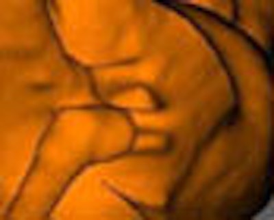
| The true-positive candidate above was a pedunculated polyp with a long stalk confirmed by both readers on axial image with 2D MPRs and 3D correlation. This polyp was also confirmed at conventional colonoscopy. Images courtesy of Dr. Mehdi Cadi. |
"The candidate polyps were listed for both prone and supine acquisition," said lead investigator Dr. Mehdi Cadi at the 2006 European Congress of Radiology (ECR) in Vienna. "Then with a single click the readers were immediately able to correlate with 2D MPR and 3D views on the same screen." The colon was divided into five segments for analysis.
 |
| Above is a false-positive polyp candidate highlighted by CAD. Both readers easily identified the finding as an ileocecal valve on axial, coronal, and 3D views. Image courtesy of Dr. Mehdi Cadi. |
Based on conventional colonoscopy results, 35% of patients (n=14) had 19 lesions 6 mm or larger, including 6 polyps larger than 10 mm, 2 of which were right colon cancers. Thirteen polyps ranged from 6-9 mm in diameter. VC's sensitivity for the detection of polyps without and with CAD were 89.4% and 94.7% for reader one, and 52.6% and 73.3% for reader two, respectively.
"CAD alone selected 195 candidates for polyps. Among them, 162 and 132 of these candidates were correctly identified as false positives by CAD by reader one and reader two," Cadi said. The majority of false positives on CAD were related to bulbous haustral folds (41%), residual stool (16%), and the ileocecal valve (9.7%), he said.
"CAD improved the performance of both readers; however, the effects of CAD were more (significant) when the reader was junior," Cadi said. "To be able to calculate the false-negative results of CAD, a correlation image by image of both classic colonoscopy and (VC) must be performed, and is still in progress," he added.
"CAD increased the sensitivity of (VC) to detect colorectal polyps," Cadi concluded. The effects of CAD are greater with beginning readers, but CAD also resulted in an improved learning curve for general radiologists, he said.
By Eric Barnes
AuntMinnie.com staff writer
May 4, 2006
Related Reading
VC CAD improves results for readers at all levels, April 7, 2006
Colon CAD helps expert readers, March 16, 2006
VC CAD found equivalent to colonoscopy in screening population, December 21, 2005
New VC reading schemes could solve old problems, October 13, 2005
Colon CAD: VC's extra eyes face new challenges, August 5, 2005
Copyright © 2006 AuntMinnie.com