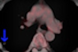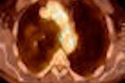
NEW YORK (Reuters Health), Oct 13 - In staging melanoma patients with palpable lymph nodes, fluorine-18 (F-18) FDG-PET and CT are equivalent, but FDG-PET detects more metastatic sites, particularly bone and subcutaneous metastases, Dutch researchers report in the October 1 issue of the Journal of Clinical Oncology.
Dr. Harald J. Hoekstra of University Medical Centre Groningen and colleagues came to these conclusions after prospectively studying data on 251 patients who underwent both FDG-PET and CT at five centers.
Distant metastases were suggested by FDG-PET in 32% of the patients and by CT in 29%. After correlation with cytology and histology or six months of follow-up, results proved correct in 27% of FDG-PET scans and 24% of CT scans.
The article notes that results were correct in 68 of 79 patients (86%) with positive FDG-PET scans and in 61 of 72 (85%) with positive CT scans.
False-positive rates were not statistically different between hospitals, according to the researchers.
Significantly more sites were detected with FDG-PET than with CT (133 versus 112). This was particularly the case for bone metastases (27 versus 10) and subcutaneous metastases (11 versus five).
Treatment changed in 19% of the patients. In most of these cases (79%), therapy was changed on the basis of both scans. Changes were made solely a result of FDG-PET in 17%, and solely a result of CT in 4%.
The researchers also calculated that FDG-PET provided value in addition to that of spiral CT in 17% of patients. Conversely, CT was of additional value in 9% of the patients.
"Due to the improved staging of melanoma patients with lymph node metastases," Dr. Hoekstra told Reuters Health, "surgical oncologists can better select melanoma patients for curative therapeutic lymph node dissection and refer patients with distant disease to a medical oncologist for systemic treatment."
Overall, he concluded, based on FDG-PET, "melanoma patients with lymph node metastases get the best tailored treatment."
By David Douglas
J Clin Oncol 2009;27:4774-4780.
Last Updated: 2009-10-12 18:00:22 -0400 (Reuters Health)
Related Reading
New melanoma imaging agent shows promise, October 1, 2009
Noncontrast CT with PET reduces melanoma radiation dose, August 12, 2009
Copyright © 2009 Reuters Limited. All rights reserved. Republication or redistribution of Reuters content, including by framing or similar means, is expressly prohibited without the prior written consent of Reuters. Reuters shall not be liable for any errors or delays in the content, or for any actions taken in reliance thereon. Reuters and the Reuters sphere logo are registered trademarks and trademarks of the Reuters group of companies around the world.