VIENNA - Diffusion-weighted imaging (DWI) is viewed increasingly as an essential and valuable tool for detecting liver metastases, but experts say they would welcome manufacturers' improvements in the signal-to-noise ratio, better image resolution and sequences, and for DWI to be less prone to artifacts. They also strongly suggest checking the capacity of new MR machines for DWI capability.
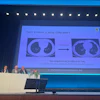
In yesterday's state-of-the-art symposium on the abdomen, Dr. Filipe Caseiro-Alves, PhD, from the University Clinic of Radiology, Coimbra University Hospitals, Portugal, discussed a series of images relating to liver and pancreatic diseases using DWI and provided examples of when the technique is most clinically useful.
"Diffusion today is an integral part of the MR protocol, and is an important component of the examination," he said. "It's an excellent method because DWI provides information on a microscopic and cellular imaging level. In the future I hope we will see more on intravoxel incoherent motion, which explores perfusion."
 Dr. Filipe Caseiro-Alves, PhD, from Coimbra, Portugal. Image courtesy of the European Society of Radiology.
Also commenting on how use of DWI could be improved, session moderator and former ECR President Dr. Yves Menu from Paris said he did not believe that all machines were equal in terms of DWI. His advice to users is to ensure that when purchasing a new MR machine, pay attention to the capacity of the machine to perform DWI.
"This might be complicated because image quality varies so much, and knowing that a machine can deliver on a regular basis every day for every patient is not possible," he noted.
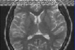
Routine clinical use of DWI began in the 1980s. The initial applications came from brain imaging for edema, as well as inflammation, among many other processes. DWI is easier to implement in the brain because it is stationary. Later, use extended to elsewhere in the body, and today it is a standard technique coupled with MRI, in particular, in abdominal examinations.
Explaining the guiding principle, Caseiro-Alves said DWI moves beyond morphology and regular MR examinations to examine structures at a microscopic level.
"DWI allows us to see how extracellular water molecules behave with Brownian motion," he said. "The technique uses radiofrequency gradients, which deposit on water molecules around the cells providing a signal intensity determined by [the degree of movement of] the water molecules."
Body fluids are not in a steady state and move randomly at a microscopic level, unless restricted by the presence of too many cells such as in a tumor. "When there are too many cells present, the water molecules are imprisoned so when you apply the sequence they provide a large signal. Other water molecules that are unrelated to the tumor do not emit a response," he explained.
Caseiro-Alves stressed that now it's mandatory to use DWI from head to toe, adding that it also had value in picking up processes that may have different cellular environments such as inflammation, for instance around small bowel. "This helps us decide if the patient needs treatment. It is more difficult to detect this on standard MRI because once inflammation is present there is difficulty in grading inflammation. However, with DWI you can grade better and more completely."
 Dr. Yves Menu from Paris. Image courtesy of the European Society of Radiology.
He also pointed out that DWI was useful for the evaluation of a tumor response to chemotherapy. If the tumor shrinks, leaving fewer cells and necrosis, then the proportion of water molecules will increase and DWI will change accordingly. "If signal intensity disappears, then this is a good reflection of treatment with the tumor disappearing. This provides the radiologist with an excellent tool to monitor patient response to therapy," he remarked.
In particular, Caseiro-Alves illustrated the usefulness of DWI for detecting metastases, which it shows very clearly. "For the first time in liver metastases, DWI shows the flowing blood in the liver as black, not as bright as usual. This means you see lesions as bright white spots against a black background," he remarked. "As such, the detection rate is high, so it is important to try DWI in the protocols for detection of pathological processes and detection of metastases."
For the pancreas, he referred to a systematic review, which concluded that use of DWI in the pancreas for solid masses did not clearly differentiate benign mass from malignancy. Specifically, he pointed out that in pancreatic cancer and autoimmune pancreatitis, the apparent diffusion coefficient (ADC) was similar. "This is logical because autoimmune pancreatitis essentially involves cellular proliferation so there will be restriction as in cancer."
Looking ahead, he said he hoped that vendors were developing ways of ameliorating the quality of the sequence, and making it less prone to artifacts so radiologists could extract more information from DWI.
"We need a higher resolution of sequences and images because this is the major problem. We can see the lesion, but we don't know exactly where it stands because the image matrix is too low and the signal-noise ratio is intrinsically poor. These signals are not the broad signal used for an MR image. We also need DWI to be less prone to artifacts," Caseiro-Alves commented.
Other presenters in the session were Dr. Harriet Thoeny from the Bern University Hospital in Switzerland, who gave a tutorial on DWI for beginners, and Dr. Sofia Gourtsoyianni from Guy's and St. Thomas' NHS Foundation Trust in London, who discussed DWI of abdominal lymph nodes.
Originally published in ECR Today on 9 March 2013.
Copyright © 2013 European Society of Radiology