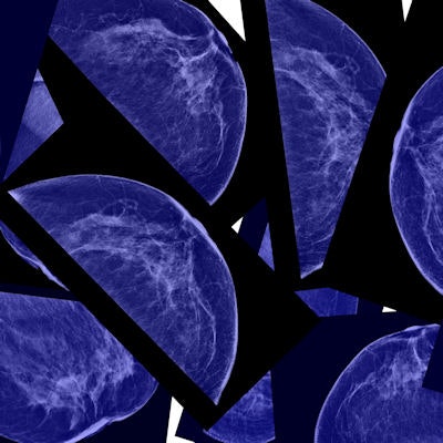
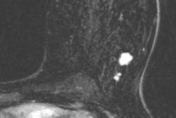
German researchers have developed a novel breast MRI technique that greatly reduces false-positive findings and increases the detection of breast cancer -- without the need for a gadolinium-based contrast agent, according to a study published online February 20 in Radiology.
The approach features a form of diffusion-weighted MRI (DWI-MRI) known as diffusion kurtosis imaging, which analyzes the distribution of water to evaluate breast tissue structures at a microscopic level. A software algorithm based on a kurtosis radiomics model then extracts imaging data to accurately characterize the lesions (Radiology, February 20, 2018).
"[The] model ... reduces false-positive results by 70% in lesions classified as BI-RADS 4 or 5 at screening mammography while retaining sensitivity greater than 98%," wrote lead author Dr. Sebastian Bickelhaupt, from the German Cancer Research Center in Heidelberg, and colleagues. "Since malignant lesions disrupt the tissue structures at this level, diffusion kurtosis might serve as a relevant marker of changes."
Contrast challenges
Previous studies have shown that with the addition of a contrast agent with ultrasound or mammography, positive predictive values can increase from 13%-17% to 69%-78% for women with more severe cases of suspected cancer, such as BI-RADS 4.
When a follow-up MRI scan is needed to better determine a diagnosis, a gadolinium-based contrast agent (GBCA) is the standard course of action. GBCAs, however, have become somewhat controversial, as studies have shown that minute traces of the element can linger in the body's tissues long after administration.
To help alleviate any concerns about gadolinium and to better manage patients who may have contraindications for contrast, the researchers developed a software algorithm based, in part, on radiomics that can gather a massive amount of data from images. They tested the algorithm in a multicenter study that analyzed patient information from DWI breast MRI exams performed between 2014 and 2016.
All of the women were referred for follow-up MRI scans and possible biopsy after a suspicious finding for cancer (BI-RADS 4 or 5) on mammography. DWI breast scans were conducted on MRI devices at two different imaging facilities. The patients underwent biopsy after the DWI scans.
One institution used a 1.5-tesla scanner (Magnetom Aera, Siemens Healthineers) and a two-channel breast coil to image 95 patients (mean age, 58.6 ± 6.6 years). In this group, 61 women had malignancies and 34 had benign lesions. This set of patients served as the training set for the kurtosis-based radiomics model. The second facility used a 1.5-tesla scanner (Ingenia, Philips Healthcare) and a two-channel breast coil to image 127 patients (mean age, 58.2 ± 6.8 years). Among these women, 61 had malignancies and 66 had benign lesions, which were used to test the model.
Enhanced detection
Among all 222 patients, histopathology results confirmed malignant lesions in 122 women (55%); invasive ductal carcinoma was the most common finding, in 90 patients (74%). Benign lesions were found in the remaining 100 women (45%); fibrosis (21 patients, 21%) and fibroadenoma (20 patients, 20%) were the most common abnormalities.
In the test group of 127 patients, the kurtosis-based radiomics model reduced the number of false-positive results from 66 to 20, achieving specificity of 70% (46 of 66). The model also detected 60 of 61 malignant lesions, for sensitivity of 98%. The model missed one true-positive result.
Breaking the results down further, the model reached sensitivity of 100% in women with BI-RADS 4A and 4B breast cancer. It also reduced false positives from 50 to 13 for the BI-RADS 4A patients and from 15 to nine for those with BI-RADS 4B cancer.
| Diagnostic performance of kurtosis radiomics model to detect lesions | ||
| Sensitivity | Specificity | |
| All patients | 98% (60/61) | 70% (46/66) |
| BI-RADS 4A | 100% (9/9) | 74% (37/50) |
| BI-RADS 4B | 100% (29/29) | 60% (9/15) |
| BI-RADS 5 | 96% (22/23) | 0% (0/1) |
"A radiomics breast cancer model based on DWI with adapted kurtosis fitting allowed for improved differentiation between malignant and benign breast lesions in both training and independent test datasets acquired by using MR imaging machines from different vendors at different institutions," Bickelhaupt and colleagues wrote.
If the findings are validated further, the researchers suggest that the kurtosis radiomics model could have additional advantages beyond reducing unnecessary biopsies in women with BI-RADS 4 lesions. They did stress, however, that their approach is not designed to replace current contrast-enhanced breast MRI protocols in general, but rather to expand diagnostic options to answer specific clinical questions.





![Examples of ultrasound findings and techniques. (A) Images in a 39-year-old male patient with a mass in the left thigh. The mass is heterogeneous on the B-mode US image (compared with the patient in D) and showed increased microvascularity (superb microvascular imaging [SMI]) and shear-wave elastography (SWE) values. Undifferentiated pleomorphic sarcoma was diagnosed at biopsy (with pleomorphic rhabdomyosarcoma in surgical specimen). (B) Images in an 18-year-old male patient with a mass in the left leg. The mass is hypoechoic on the B-mode image, with no other findings suggestive of malignancy. The lesion is in contact with the cortex of the tibia, which is slightly irregular. CT revealed a doubtful anteromedial tibial erosion. The microvascular study demonstrated high vascularization, suggestive of malignancy. Periosteal Ewing sarcoma was diagnosed with both histologic and immunohistochemical confirmation. (C) Images in a 69-year-old female patient with a lump growing on the outside of the left leg. Multiple SWE examinations were performed (please note the high values obtained in the measurements, whereas the color map highlights the stiffness relative to adjacent tissues). SMI showed areas of increased vascularization to target for sampling. Undifferentiated spindle cell sarcoma was diagnosed at biopsy, with residual leiomyosarcoma in the surgical specimen after neoadjuvant therapy. (D) Images in a 56-year-old female patient with a mass in the right thigh. The mass is heterogeneous at both B-mode ultrasound (similar to patient A) and MRI (coronal T2-weighted spectral attenuated inversion recovery [SPAIR]; T1-weighted pre-contrast and postcontrast imaging), which even shows uptake after the administration of paramagnetic contrast material, which is traditionally suggestive of malignancy. Low values at SMI and elastography are suggestive of benignity. Spindle cell lipoma was diagnosed at biopsy, with atypical spindle cell lipomatous tumor in the surgical specimen.](https://img.auntminnieeurope.com/mindful/smg/workspaces/default/uploads/2026/08/images-radiol250278fig2.APCFLSvX6p.jpg?auto=format%2Ccompress&dpr=2&fit=crop&h=167&q=70&w=250)


