Preliminary results are encouraging from a Swiss clinical trial of a novel PET/CT radiotracer being investigated for visualizing tumors in patients with esophageal cancer.
A group led by Dr. Matthieu Dietz of Lausanne University Hospital found that while standard F-18 FDG-PET/CT imaging detected more total tumors, the new approach enabled the visualization of angiogenesis, the process through which tumors develop blood supplies to grow. The study findings were published online on 1 February in the European Journal of Hybrid Imaging.
"Ga-68 NODAGA-RGD-PET/CT is a noninvasive, holistic imaging of tumor angiogenesis and could play a pivotal role in identifying patients which have greatest benefit from anti-angiogenic therapy," the authors wrote.
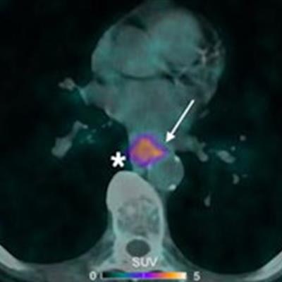
 Ga-68 NODAGA-RGD-PET/CT (A), PET (B, D) and CT (C) and F-18 FDG-PET/CT (E), PET (F, H) and CT (G) axial views of a primary lesion (asterisks) in a 53-year-old participant with esophageal squamous cell carcinoma. Note intense homogenous uptake in the primary lesion on F-18 FDG-PET/CT (SUVmax 12.1 g/mL, metabolic tumor volume 2.54 cm3), whereas the primary lesion demonstrates a different uptake pattern in the corresponding Ga-68 NODAGA-RGD-PET/CT image: a weaker and more heterogeneous uptake (SUVmax 5.1 g/mL), seen mostly in the periphery of the tumor, with a slight extension in perilesional structures (arrows), and a larger metabolic tumor volume (metabolic tumor volume 5.35 cm3). Image courtesy of the European Journal of Hybrid Imaging through CC BY 4.0.
Esophageal cancer is the seventh most common cancer worldwide and accounts for more than half a million deaths each year. F-18 FDG-PET/CT detects tumors in these patients by visualizing abnormal tumor glucose metabolism, yet Ga-68 NODAGA-RGD PET/CT targets different tumor activity, the researchers explained.
Ga-68 NODAGA-RGD is made of a peptide ligand (arginine-glycine-aspartate, or RDG) that binds to integrin receptors (avb3) on endothelial cells. These cells are overactive in cancer and involved in forming vessels that supply blood to tumors. Combined with a gallium-68 (Ga-68) isotope, the tracer visualizes this activity on PET. Numerous such PET tracers have been developed, with this being the first time one has been tested in patients with esophageal cancers, noted Dietz, who took up a post at Hôpital Louis Pradel, Lyon, France, in November 2022.
Trial logistics
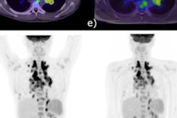
The group performed 10 F-18 FDG-PET/CT scans and 10 Ga-68 NODAGA-RGD-PET/CT scans (Discovery 690, GE HealthCare) in nine patients with biopsy-confirmed esophageal or gastroesophageal junction cancer (EGJ) enrolled in the trial. The time between scans ranged from one to nine days.
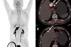
According to the findings, Ga-68 NODAGA-RGD-PET/CT detected positive radiotracer uptake in 10 primary sites (eight for primary tumors and two for local relapse suspicion), six lymph nodes, and three skeletal sites. F-18 FDG-PET/CT detected positive uptake in the same sites but also in 16 additional lymph nodes and one adrenal gland.
However, the researchers noted that while F-18 FDG uptake was homogenous inside all the confirmed primary sites (n = 9), Ga-68 NODAGA-RGD showed more heterogenous uptake in six out of the nine confirmed primary sites (67%). These sites were involved in potential early angiogenesis, with activity on imaging seen mostly in the periphery of the tumor and extending into surrounding tissue in five out of the nine confirmed primary sites (56%), they wrote.
"The results suggest that PET imaging of integrin avb3 expression may provide complementary information and could aid in tumor diversity and delineation," the researchers wrote.
The researchers noted that the small number of patients limited the study's statistical power and that the study is ongoing.
Ultimately, F-18 FDG-PET/CT is widely accepted as the preferred method for initial tumor staging in esophageal cancer. The extension of Ga-68 NODAGA-RGD tracer uptake into tissue around the tumors seen in this study bodes well for efforts to visualize early angiogenesis, which is a significant target for new therapies, the group wrote.
"We strongly believe that the complementary information provided by molecular imaging of avb3 expression could be clinically relevant," Dietz and colleagues concluded.