German clinicians have reported finding a rare tenosynovial giant cell tumor (TGCT) in a COVID-19 patient's thigh using gallium-68 (Ga-68) fibroblast activation protein inhibitor (FAPI) PET imaging, according to a case published on 26 January in the Journal of Nuclear Medicine.
The incidental detection highlights the high sensitivity of FAPI-PET for detecting previously unknown tumors, noted a team led by Dr. Timur Sellmann, head of anesthesiology and intensive medicine at the Evangelical Hospital Bethesda in Duisburg.
"Pathologic reassessment showed the definite diagnosis of a tenosynovial giant cell tumor, which is absolutely rare in this location," the group wrote.
TGCT is a rare type of soft tissue tumor that forms in the joints and although it is not typically cancerous, it can grow and damage surrounding structures, Sellmann and colleagues wrote. TGCTs are completely distinct from and should not be confused with any giant cell-rich tumors of the bone or soft tissue, they noted.
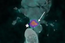
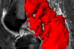
Ga-68 FAPI-PET/CT imaging in a 55-year-old male patient. No increased pulmonary tracer uptake was found (A, C). Tracer uptake colocalized with a sharply circumscribed, inhomogeneously contrast-enhancing mass on MRI without relevant diffusion restriction (B). Pathologic assessment diagnosed a tenosynovial giant cell tumor (D). Image courtesy of the Journal of Nuclear Medicine.Fibroblast activation protein (FAP) is overexpressed by cancer cells, and researchers suspect it is an essential component driving the growth of tumors. Experimental FAP-targeted molecular imaging radiotracers, such as FAPI-04 and FAPI-46, have shown promise in cancer research as well as for visualizing myocardial fibrosis.
Sellmann's team reported the rare tumor finding in a 55-year-old patient who underwent imaging due to persistent pulmonary symptoms after recovery from COVID-19. The patient was enrolled in a first-in-human trial testing Ga-68 FAPI-04 PET/CT scans (Biograph mCT, Siemens Healthineers) for identifying disease progression in 21 patients with interstitial lung disease.
The trial showed no increase in pulmonary radiotracer, but the researchers observed focally increased Ga-68 FAPI uptake in a mass in the patient's left thigh. Surgeons performed complete resection of the tumor.
"This report demonstrates the high sensitivity of Ga-68 FAPI-PET/CT to detect previously unknown tumors, making it a promising test in cases of suspected tumor disease or in cancers of unknown primary," the group concluded.


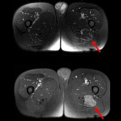
![Examples of ultrasound findings and techniques. (A) Images in a 39-year-old male patient with a mass in the left thigh. The mass is heterogeneous on the B-mode US image (compared with the patient in D) and showed increased microvascularity (superb microvascular imaging [SMI]) and shear-wave elastography (SWE) values. Undifferentiated pleomorphic sarcoma was diagnosed at biopsy (with pleomorphic rhabdomyosarcoma in surgical specimen). (B) Images in an 18-year-old male patient with a mass in the left leg. The mass is hypoechoic on the B-mode image, with no other findings suggestive of malignancy. The lesion is in contact with the cortex of the tibia, which is slightly irregular. CT revealed a doubtful anteromedial tibial erosion. The microvascular study demonstrated high vascularization, suggestive of malignancy. Periosteal Ewing sarcoma was diagnosed with both histologic and immunohistochemical confirmation. (C) Images in a 69-year-old female patient with a lump growing on the outside of the left leg. Multiple SWE examinations were performed (please note the high values obtained in the measurements, whereas the color map highlights the stiffness relative to adjacent tissues). SMI showed areas of increased vascularization to target for sampling. Undifferentiated spindle cell sarcoma was diagnosed at biopsy, with residual leiomyosarcoma in the surgical specimen after neoadjuvant therapy. (D) Images in a 56-year-old female patient with a mass in the right thigh. The mass is heterogeneous at both B-mode ultrasound (similar to patient A) and MRI (coronal T2-weighted spectral attenuated inversion recovery [SPAIR]; T1-weighted pre-contrast and postcontrast imaging), which even shows uptake after the administration of paramagnetic contrast material, which is traditionally suggestive of malignancy. Low values at SMI and elastography are suggestive of benignity. Spindle cell lipoma was diagnosed at biopsy, with atypical spindle cell lipomatous tumor in the surgical specimen.](https://img.auntminnieeurope.com/mindful/smg/workspaces/default/uploads/2026/08/images-radiol250278fig2.APCFLSvX6p.jpg?auto=format%2Ccompress&fit=crop&h=100&q=70&w=100)


![Examples of ultrasound findings and techniques. (A) Images in a 39-year-old male patient with a mass in the left thigh. The mass is heterogeneous on the B-mode US image (compared with the patient in D) and showed increased microvascularity (superb microvascular imaging [SMI]) and shear-wave elastography (SWE) values. Undifferentiated pleomorphic sarcoma was diagnosed at biopsy (with pleomorphic rhabdomyosarcoma in surgical specimen). (B) Images in an 18-year-old male patient with a mass in the left leg. The mass is hypoechoic on the B-mode image, with no other findings suggestive of malignancy. The lesion is in contact with the cortex of the tibia, which is slightly irregular. CT revealed a doubtful anteromedial tibial erosion. The microvascular study demonstrated high vascularization, suggestive of malignancy. Periosteal Ewing sarcoma was diagnosed with both histologic and immunohistochemical confirmation. (C) Images in a 69-year-old female patient with a lump growing on the outside of the left leg. Multiple SWE examinations were performed (please note the high values obtained in the measurements, whereas the color map highlights the stiffness relative to adjacent tissues). SMI showed areas of increased vascularization to target for sampling. Undifferentiated spindle cell sarcoma was diagnosed at biopsy, with residual leiomyosarcoma in the surgical specimen after neoadjuvant therapy. (D) Images in a 56-year-old female patient with a mass in the right thigh. The mass is heterogeneous at both B-mode ultrasound (similar to patient A) and MRI (coronal T2-weighted spectral attenuated inversion recovery [SPAIR]; T1-weighted pre-contrast and postcontrast imaging), which even shows uptake after the administration of paramagnetic contrast material, which is traditionally suggestive of malignancy. Low values at SMI and elastography are suggestive of benignity. Spindle cell lipoma was diagnosed at biopsy, with atypical spindle cell lipomatous tumor in the surgical specimen.](https://img.auntminnieeurope.com/mindful/smg/workspaces/default/uploads/2026/08/images-radiol250278fig2.APCFLSvX6p.jpg?auto=format%2Ccompress&fit=crop&h=112&q=70&w=112)