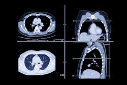
Brainomix’s e-Lung AI imaging software has been selected by Boehringer Ingelheim to provide objective, quantitative high-resolution CT scan (HRCT) imaging biomarkers as a co-primary endpoint in a phase III study for interstitial lung disease (ILD).
The DROP-FPF trial is a phase IIIB double-blind, randomized, placebo-controlled study investigating the safety and effectiveness of nerandomilast (Jascayd) in people with interstitial lung abnormalities and a family history of pulmonary fibrosis. It is the first phase III trial to use automated, quantitative HRCT imaging biomarkers, according to Brainomix.
The study, which is set to begin enrolling patients in January, will investigate whether early intervention with nerandomilast slows the progression of early signs of pulmonary fibrosis in people with a family history of the condition. It will include a two-year follow-up period, according to Brainomix.
Nerandomilast received U.S. Food and Drug Administration (FDA) approval for both idiopathic pulmonary fibrosis and progressive pulmonary fibrosis in December 2025.
Trained on large and diverse datasets of patients with different forms of ILD, e-Lung software was validated in the phase III INBUILD study, which led to the approval of nintedanib (Ofev) for patients with progressive pulmonary fibrosis, the firm added.



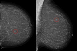
![A normal mammogram confirmed by three-year radiologic follow-up illustrates reader-marked regions of interest (ROIs) during (A) unaided (round 1) and (B) artificial intelligence (AI)–assisted (round 2) reading. Each colored dot represents an ROI for recall by a human reader. Readers could mark more than one ROI per case, represented by multiple dots of the same color. During AI-assisted reading, the AI system displayed three visible prompts: two with suspicion of malignancy scores of 35% (left mediolateral oblique [L MLO] and craniocaudal [L CC]) and one with a suspicion of malignancy score of 10% (right craniocaudal [R CC]), shown as polygonal overlays. Without AI, six of 10 readers (60%) marked a false-positive ROI. With AI assistance, this fell to two of 10 (20%). R MLO = right mediolateral oblique.](https://img.auntminnieeurope.com/mindful/smg/workspaces/default/uploads/2026/07/2026-07-14-radiology-mammogram-ai-auto-bias.H0bYO8QlWs.jpg?auto=format%2Ccompress&fit=crop&h=100&q=70&w=100)


![A normal mammogram confirmed by three-year radiologic follow-up illustrates reader-marked regions of interest (ROIs) during (A) unaided (round 1) and (B) artificial intelligence (AI)–assisted (round 2) reading. Each colored dot represents an ROI for recall by a human reader. Readers could mark more than one ROI per case, represented by multiple dots of the same color. During AI-assisted reading, the AI system displayed three visible prompts: two with suspicion of malignancy scores of 35% (left mediolateral oblique [L MLO] and craniocaudal [L CC]) and one with a suspicion of malignancy score of 10% (right craniocaudal [R CC]), shown as polygonal overlays. Without AI, six of 10 readers (60%) marked a false-positive ROI. With AI assistance, this fell to two of 10 (20%). R MLO = right mediolateral oblique.](https://img.auntminnieeurope.com/mindful/smg/workspaces/default/uploads/2026/07/2026-07-14-radiology-mammogram-ai-auto-bias.H0bYO8QlWs.jpg?auto=format%2Ccompress&fit=crop&h=112&q=70&w=112)