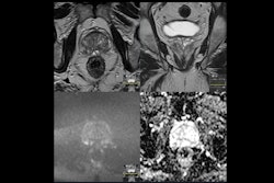
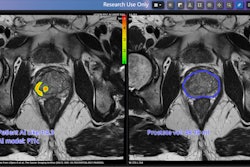
Lucida Medical and Hampshire Hospitals NHS Foundation Trust in the U.K. are highlighting results from a clinical study regarding Lucida's Prostate Intelligence (Pi) AI software for prostate cancer detection.
The five-year study, called Prostate AI Research -1 (PAIR-1), has shown that the software -- in use in NHS and European hospitals -- demonstrates performance equivalent to that of expert radiologists for identifying the disease.
PAIR-1 is a collaboration between Lucida Medical and eight NHS Trusts and has included data from more than 2,000 patients to develop, train, and validate the software, which analyzes MRI scans for clinically significant prostate cancer.
Results from the research were presented by Francesco Giganti of University College London at the recent ECR meeting in Vienna. They were also published 28 February in the European Journal of Radiology.
"[Our] research found that Pi is non-inferior to multidisciplinary team-supported radiologists across a validation set of sequential cases from six NHS hospitals with a wide range of MRI scanner types," Giganti said in a statement released by the firm. "This is the first time that a commercial AI for prostate MRI has been tested on diverse, real-world data."




![A normal mammogram confirmed by three-year radiologic follow-up illustrates reader-marked regions of interest (ROIs) during (A) unaided (round 1) and (B) artificial intelligence (AI)–assisted (round 2) reading. Each colored dot represents an ROI for recall by a human reader. Readers could mark more than one ROI per case, represented by multiple dots of the same color. During AI-assisted reading, the AI system displayed three visible prompts: two with suspicion of malignancy scores of 35% (left mediolateral oblique [L MLO] and craniocaudal [L CC]) and one with a suspicion of malignancy score of 10% (right craniocaudal [R CC]), shown as polygonal overlays. Without AI, six of 10 readers (60%) marked a false-positive ROI. With AI assistance, this fell to two of 10 (20%). R MLO = right mediolateral oblique.](https://img.auntminnieeurope.com/mindful/smg/workspaces/default/uploads/2026/07/2026-07-14-radiology-mammogram-ai-auto-bias.H0bYO8QlWs.jpg?auto=format%2Ccompress&fit=crop&h=100&q=70&w=100)


![A normal mammogram confirmed by three-year radiologic follow-up illustrates reader-marked regions of interest (ROIs) during (A) unaided (round 1) and (B) artificial intelligence (AI)–assisted (round 2) reading. Each colored dot represents an ROI for recall by a human reader. Readers could mark more than one ROI per case, represented by multiple dots of the same color. During AI-assisted reading, the AI system displayed three visible prompts: two with suspicion of malignancy scores of 35% (left mediolateral oblique [L MLO] and craniocaudal [L CC]) and one with a suspicion of malignancy score of 10% (right craniocaudal [R CC]), shown as polygonal overlays. Without AI, six of 10 readers (60%) marked a false-positive ROI. With AI assistance, this fell to two of 10 (20%). R MLO = right mediolateral oblique.](https://img.auntminnieeurope.com/mindful/smg/workspaces/default/uploads/2026/07/2026-07-14-radiology-mammogram-ai-auto-bias.H0bYO8QlWs.jpg?auto=format%2Ccompress&fit=crop&h=112&q=70&w=112)