Researchers from Amsterdam University Medical Center have developed an AI algorithm for reading FDG-PET/CT scans that performs better than standard clinical scores used to identify high-risk lymphoma patients.
A group led by doctoral candidate Maria Ferrandez trained a convolutional neural network (CNN) on F-18 FDG-PET/CT baseline scans of patients with diffuse large B-cell lymphoma (DLBCL). The model was highly accurate at predicting the probability of disease progression for patients within two years, the study authors found.
"The CNN model predicted two-year time-to-progression in DLBCL patients better than IPI [International Prognostic Index] scores," the group wrote in an article published on 12 August in Scientific Reports.
IPI scores include a number of clinical metrics and are used by clinicians to determine the number of treatment cycles patients with lymphoma may undergo. Overall, however, one-third of DLBCL patients assessed using these scores do not respond to first-line treatment, the team noted.
F-18 FDG-PET/CT is a valuable approach for characterizing the severity of lymphoma tumors and is frequently used as an early outcome prediction marker, the group added. Thus, the researchers aimed to develop an AI algorithm using such scans from DLBCL patients that could help identify high-risk patients more effectively.
The group collected 296 baseline scans from patients in a previous prospective clinical trial to test treatment with rituximab plus cyclophosphamide, doxorubicin, vincristine, and prednisone (so-called "R-CHOP" cycles). Baseline maximum intensity projections (MIPs) were used to train the CNN model, and a separate external dataset of images from 340 DLBCL patients served to validate the model.
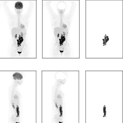
 Illustration of the different maximum intensity projections (MIPs) implemented in the study. (a) Coronal view. (b) Sagittal view. From left to right: MIP, MIP without brain, and lesion MIP. Image and caption courtesy of Scientific Reports through CC BY 4.0.
The CNN provided a two-year time-to-progression prediction with an area under the curve (AUC) of 0.74 and outperformed the IPI-based model (AUC = 0.68). In addition, the researchers synthetically removed the tumors from the FDG-PET/CT scans and ran the CNN again on the R-CHOP dataset. In these cases, high probabilities (> 0.6) predicted by the CNN were considerably decreased, they found.
"It was illustrated that the model prediction is affected by the presence or absence of lesions," the researchers wrote.
Ultimately, this study was meant to be a first step in the exploratory analysis of using end-to-end CNNs in lymphoma patients, according to the researchers.
"Even though further investigations are necessary, our current findings suggest that CNNs using MIPs have potential as outcome prediction models," they concluded.
The full article is available here.