Helsinki-based Mvision AI has released new software, version 1.2.4, for the company's guideline-based automation segmentation service. The vendor's contouring product is also getting a rebranding.
Contour+ is a component of Mvision AI's Guideline-Based Segmentation platform. The platform provides automation, verification, and continuous education for the introduction of AI contouring into medical department. Contour+ is CE-marked and has been granted 510(k) clearance by the U.S. Food and Drug Administration (FDA).
Mvision AI said Contour+ has been updated with new models to serve clinical needs. These include the following:
- A new male pelvis T2-weighted MR model supporting MR-only clinical workflow and MR-based contouring and planning is now available.
- A brain T1-weighted MR model with new scanner data for performance improvement and MR-based contouring is now available.
- Abdomen and breast models have been updated to include heart sub-structures for cardiac toxicity assessment or personalized plan optimization. RTOG & RADCOMP guideline style for breasts and lymph nodes has also been added.
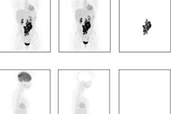
- A new whole-body model is available to boost contouring speed for patients being treated for multiple localizations.
- Female pelvis model now includes the lymph node volumes and other clinically meaningful structures to support clinicians with their target contouring.
- A new bone CT model contouring independent vertebrae and ribs is also now available.
Contour+ has an industry standard-compliant AI development process that follows international consensus contouring guidelines.




![A normal mammogram confirmed by three-year radiologic follow-up illustrates reader-marked regions of interest (ROIs) during (A) unaided (round 1) and (B) artificial intelligence (AI)–assisted (round 2) reading. Each colored dot represents an ROI for recall by a human reader. Readers could mark more than one ROI per case, represented by multiple dots of the same color. During AI-assisted reading, the AI system displayed three visible prompts: two with suspicion of malignancy scores of 35% (left mediolateral oblique [L MLO] and craniocaudal [L CC]) and one with a suspicion of malignancy score of 10% (right craniocaudal [R CC]), shown as polygonal overlays. Without AI, six of 10 readers (60%) marked a false-positive ROI. With AI assistance, this fell to two of 10 (20%). R MLO = right mediolateral oblique.](https://img.auntminnieeurope.com/mindful/smg/workspaces/default/uploads/2026/07/2026-07-14-radiology-mammogram-ai-auto-bias.H0bYO8QlWs.jpg?auto=format%2Ccompress&fit=crop&h=100&q=70&w=100)


![A normal mammogram confirmed by three-year radiologic follow-up illustrates reader-marked regions of interest (ROIs) during (A) unaided (round 1) and (B) artificial intelligence (AI)–assisted (round 2) reading. Each colored dot represents an ROI for recall by a human reader. Readers could mark more than one ROI per case, represented by multiple dots of the same color. During AI-assisted reading, the AI system displayed three visible prompts: two with suspicion of malignancy scores of 35% (left mediolateral oblique [L MLO] and craniocaudal [L CC]) and one with a suspicion of malignancy score of 10% (right craniocaudal [R CC]), shown as polygonal overlays. Without AI, six of 10 readers (60%) marked a false-positive ROI. With AI assistance, this fell to two of 10 (20%). R MLO = right mediolateral oblique.](https://img.auntminnieeurope.com/mindful/smg/workspaces/default/uploads/2026/07/2026-07-14-radiology-mammogram-ai-auto-bias.H0bYO8QlWs.jpg?auto=format%2Ccompress&fit=crop&h=112&q=70&w=112)