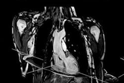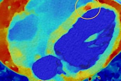
Royal Philips, the parent of Philips Healthcare, announced it will implement a regional data integration hub for the U.K.'s National COVID-19 Chest Imaging Database (NCCID), a resource of x-ray, CT, and MRI images from COVID-19 hospital patients across the country.
Primarily, the database is being used for the validation of artificial intelligence (AI) products for use in the National Health Service (NHS), which the organizations hope will help in the diagnosis and treatment of COVID-19 in the U.K. The database of clinical images has the potential to enable quicker assessments of patients when they arrive at hospitals' emergency departments, increase the safety and consistency of care across the country, and ultimately save lives.
The database is being made available to researchers, clinicians, technology companies, and all those wanting to investigate the disease and develop solutions that can support the COVID-19 patient-care pathway.
The NCCID database was developed in collaboration with NHXS, a company involved in the digital transformation of the NHS, the British Society of Thoracic Imaging, and the Royal Surrey NHS Foundation Trust.




![A normal mammogram confirmed by three-year radiologic follow-up illustrates reader-marked regions of interest (ROIs) during (A) unaided (round 1) and (B) artificial intelligence (AI)–assisted (round 2) reading. Each colored dot represents an ROI for recall by a human reader. Readers could mark more than one ROI per case, represented by multiple dots of the same color. During AI-assisted reading, the AI system displayed three visible prompts: two with suspicion of malignancy scores of 35% (left mediolateral oblique [L MLO] and craniocaudal [L CC]) and one with a suspicion of malignancy score of 10% (right craniocaudal [R CC]), shown as polygonal overlays. Without AI, six of 10 readers (60%) marked a false-positive ROI. With AI assistance, this fell to two of 10 (20%). R MLO = right mediolateral oblique.](https://img.auntminnieeurope.com/mindful/smg/workspaces/default/uploads/2026/07/2026-07-14-radiology-mammogram-ai-auto-bias.H0bYO8QlWs.jpg?auto=format%2Ccompress&fit=crop&h=100&q=70&w=100)


![A normal mammogram confirmed by three-year radiologic follow-up illustrates reader-marked regions of interest (ROIs) during (A) unaided (round 1) and (B) artificial intelligence (AI)–assisted (round 2) reading. Each colored dot represents an ROI for recall by a human reader. Readers could mark more than one ROI per case, represented by multiple dots of the same color. During AI-assisted reading, the AI system displayed three visible prompts: two with suspicion of malignancy scores of 35% (left mediolateral oblique [L MLO] and craniocaudal [L CC]) and one with a suspicion of malignancy score of 10% (right craniocaudal [R CC]), shown as polygonal overlays. Without AI, six of 10 readers (60%) marked a false-positive ROI. With AI assistance, this fell to two of 10 (20%). R MLO = right mediolateral oblique.](https://img.auntminnieeurope.com/mindful/smg/workspaces/default/uploads/2026/07/2026-07-14-radiology-mammogram-ai-auto-bias.H0bYO8QlWs.jpg?auto=format%2Ccompress&fit=crop&h=112&q=70&w=112)