Royal Philips and the Spanish National Center for Cardiovascular Research have developed an MRI technique that provides a complete cardiac study in less than a minute.
The MRI protocol is able to acquire images that visualize the shape and function of the heart, all in approximately 20 seconds. Another 20 seconds is needed to evaluate the degree of fibrous after cardiac muscle death, according to Philips. Typically, this process takes about one hour.
The new technique, which is called enhanced SENSE by static outer-volume subtraction (ESSOS), is based on the fact that -- except for the heart -- a patient's chest anatomy remains stationary during a breath-hold exam.
With ESSOS, after an initial image of the static, outer volume of the heart is captured, this MRI data is temporarily removed. Since the MRI signal of the beating heart can more easily be subtracted from the subsequent scan data, acquiring a 3D image of the heart can be performed four times faster. After the dynamic of the beating heart is reconstructed, the static outer volume images are returned to generate a full 3D cardiac image.
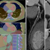
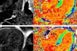
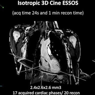
 The new ESSOS cardiac MRI protocol can acquire faster 3D images. Image courtesy of Philips.
More than 100 patients with heart issues participated in a clinical trial in which both the conventional and the ESSOS method were used. Radiologists determined that both image types were in excellent agreement on heart function measurements and the characterization of tissue damage to a patient's heart muscle, according to trial results published in April 2021 in the Journal of the American College of Cardiology.
Additionally, the method can be used with existing phased-array MRI scanners without modification.