
QT Imaging and software developer Olea Medical are collaborating to bolster QT Imaging’s breast imaging platform.
Under the agreement, Olea Medical’s AI-ready imaging technology will be integrated into QT Imaging’s Cloud SaaS Platform for secure cloud-based delivery of QT Imaging Breast Acoustic CT studies incorporating Olea Medical’s validated visualization, advanced quantitative analysis technology, and multimodality integration, the firms said.
QT Imaging will distribute the QT Imaging Olea Vision software, a customizable 3D and 4D DICOM viewer with user-defined hanging protocols and intuitive navigation across DICOM series, advanced 3D data loading and visualization, and image manipulation.
The combined tool will support a scalable multimodality breast imaging approach, integrating mammography, digital breast tomosynthesis (DBT), ultrasound, MR imaging, and QT Imaging studies. The software also includes subtraction tools to support enhanced diagnostic assessment, the partners added.
Other features include secure, network-enabled access enabling multi-institutional collaboration, centralized research workflows, and standardized clinical trial support, according to QT Imaging and Olea Medical.





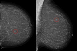



![A normal mammogram confirmed by three-year radiologic follow-up illustrates reader-marked regions of interest (ROIs) during (A) unaided (round 1) and (B) artificial intelligence (AI)–assisted (round 2) reading. Each colored dot represents an ROI for recall by a human reader. Readers could mark more than one ROI per case, represented by multiple dots of the same color. During AI-assisted reading, the AI system displayed three visible prompts: two with suspicion of malignancy scores of 35% (left mediolateral oblique [L MLO] and craniocaudal [L CC]) and one with a suspicion of malignancy score of 10% (right craniocaudal [R CC]), shown as polygonal overlays. Without AI, six of 10 readers (60%) marked a false-positive ROI. With AI assistance, this fell to two of 10 (20%). R MLO = right mediolateral oblique.](https://img.auntminnieeurope.com/mindful/smg/workspaces/default/uploads/2026/07/2026-07-14-radiology-mammogram-ai-auto-bias.H0bYO8QlWs.jpg?auto=format%2Ccompress&dpr=2&fit=crop&h=167&q=70&w=250)


