
New guidelines jointly developed by multiple Italian medical societies call for AI to decide which mammograms should go to single or double reading by radiologists.
The guidelines were developed by 10 scientific societies. The effort was coordinated by the National Screening Observatory, and contributions were made by the Centre for Research on Health and Social Care Management (CERGAS) and the F. Dondena Center at Bocconi University.
The authors noted that AI can support image selection, which can help reduce fatigue and diagnostic variability. They also highlighted the potential for better distribution of workloads in clinics facing shortages of radiologists and better facilitation for extending screening programs in regions with greater operational difficulties.
The guidelines also incorporate two other recommendations already included in the most recent European Guidelines. The first suggests integrating AI after an initial positive human opinion to increase the likelihood of detecting lesions at an early stage. The second recommends not replacing the second radiologist with AI, believing that the technologies are not yet mature enough for such widespread use.
The recommendations are defined as nonbinding and addressed to both healthcare professionals and regional institutions.
The following group developed the guidelines: Italian Association of Epidemiology (AIE), Italian Association of Medical Physics (AIFM), Italian Association of Medical Oncology (AIOM), Italian Association of Radiotherapy and Clinical Oncology (AIRO), FASTeR/AITERS, Italian Mammography Screening Group (GISMa), Italian Society of Pathological Anatomical and Diagnostic Cytology (SIAPEC-IAP), Italian Society of Plastic, Reconstructive and Aesthetic Surgery (SICPRE), Italian Society of Medical and Interventional Radiology (SIRM), and Italian Society of Hygiene, Preventive Medicine and Public Health (SITI).





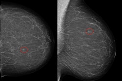



![A normal mammogram confirmed by three-year radiologic follow-up illustrates reader-marked regions of interest (ROIs) during (A) unaided (round 1) and (B) artificial intelligence (AI)–assisted (round 2) reading. Each colored dot represents an ROI for recall by a human reader. Readers could mark more than one ROI per case, represented by multiple dots of the same color. During AI-assisted reading, the AI system displayed three visible prompts: two with suspicion of malignancy scores of 35% (left mediolateral oblique [L MLO] and craniocaudal [L CC]) and one with a suspicion of malignancy score of 10% (right craniocaudal [R CC]), shown as polygonal overlays. Without AI, six of 10 readers (60%) marked a false-positive ROI. With AI assistance, this fell to two of 10 (20%). R MLO = right mediolateral oblique.](https://img.auntminnieeurope.com/mindful/smg/workspaces/default/uploads/2026/07/2026-07-14-radiology-mammogram-ai-auto-bias.H0bYO8QlWs.jpg?auto=format%2Ccompress&dpr=2&fit=crop&h=167&q=70&w=250)


