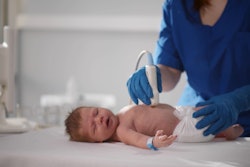
An intensive training program helped improve ultrasound acquisition skills for midwives scanning pregnant women, according to a study published on 16 July in WFUMB Ultrasound Open.
A research group led by radiographer Shown Haluzani from Lusaka Apex Medical University in Zambia described the success of the training program, which translated to higher competency and good image quality from participating midwives.
“These findings are essential for guiding interventions aimed at enhancing ultrasound service delivery and ensuring consistent, high-quality healthcare outcomes because this is critical for effective prenatal care in Zambia,” Haluzani and colleagues wrote. “This has high potential to improve prenatal care.”
Limited access to ultrasound services in low and middle-income countries makes it necessary to develop intensive training programs for health professionals, including midwives. Previous studies have highlighted the success of training programs around the world in training non-imaging professionals on using ultrasound for pregnancy scans: One team described their experience working with sonographers in Nepal on point-of-care ultrasound (POCUS) in pregnant women, while another reported the success of a POCUS training program in Haiti, which showed improved performance among physicians.
The Haluzani group evaluated the quality of ultrasound images produced by midwives, assessing their competency in gestational age assessment using femur length. It conducted the study in 11 healthcare facilities across four Zambian districts, including 928 images captured during implementation of the Training in Ultrasound to Determine Gestational Age (TUDA) program in 2021. The program trained 24 midwives for 14 days, followed by eight weeks of supervised scanning.
The midwives achieved a competency level of 97.95% in basic ultrasound scans for determining gestational age using femur length. They also achieved an average proportion of good-quality images at 87.95% as assessed by four evaluators. Haluzani and colleagues did not find statistically significant differences among assessors, indicating consistency in evaluations.
“Trainers and radiographers exhibited different average proportions of good-quality images at 91.5% and 83.9%, respectively, with no statistically significant differences observed between them,” they wrote.
However, the group did find significant variation in image quality among different facilities. In the Chipata facility, 24.32% of scans were classified as poor quality, but at George Clinic, only 2.3% were considered poor quality. Through a Chi-square test, the team found a substantial correlation between health facility and image quality (χ2 = 15.99, p = 0.025) but observed no significant correlations between image quality and factors such as work experience, age group, or gender.
In the end, the study results provide “critical evidence” for successfully training midwives to produce high-quality ultrasound images in this area, according to the investigators.
“High-quality ultrasound images allow for consistent interpretation by various observers in making a diagnosis which will assist in determination of a positive pregnancy outcome,” they wrote.
Finally, Haluzani et al recommended including ultrasound scanning in-service and preservice training, as well as establishing standardized protocols and guidelines for ultrasound scanning services during antenatal clinics.
The study can be found in its entirety here.

![Examples of ultrasound findings and techniques. (A) Images in a 39-year-old male patient with a mass in the left thigh. The mass is heterogeneous on the B-mode US image (compared with the patient in D) and showed increased microvascularity (superb microvascular imaging [SMI]) and shear-wave elastography (SWE) values. Undifferentiated pleomorphic sarcoma was diagnosed at biopsy (with pleomorphic rhabdomyosarcoma in surgical specimen). (B) Images in an 18-year-old male patient with a mass in the left leg. The mass is hypoechoic on the B-mode image, with no other findings suggestive of malignancy. The lesion is in contact with the cortex of the tibia, which is slightly irregular. CT revealed a doubtful anteromedial tibial erosion. The microvascular study demonstrated high vascularization, suggestive of malignancy. Periosteal Ewing sarcoma was diagnosed with both histologic and immunohistochemical confirmation. (C) Images in a 69-year-old female patient with a lump growing on the outside of the left leg. Multiple SWE examinations were performed (please note the high values obtained in the measurements, whereas the color map highlights the stiffness relative to adjacent tissues). SMI showed areas of increased vascularization to target for sampling. Undifferentiated spindle cell sarcoma was diagnosed at biopsy, with residual leiomyosarcoma in the surgical specimen after neoadjuvant therapy. (D) Images in a 56-year-old female patient with a mass in the right thigh. The mass is heterogeneous at both B-mode ultrasound (similar to patient A) and MRI (coronal T2-weighted spectral attenuated inversion recovery [SPAIR]; T1-weighted pre-contrast and postcontrast imaging), which even shows uptake after the administration of paramagnetic contrast material, which is traditionally suggestive of malignancy. Low values at SMI and elastography are suggestive of benignity. Spindle cell lipoma was diagnosed at biopsy, with atypical spindle cell lipomatous tumor in the surgical specimen.](https://img.auntminnieeurope.com/mindful/smg/workspaces/default/uploads/2026/08/images-radiol250278fig2.APCFLSvX6p.jpg?auto=format%2Ccompress&fit=crop&h=100&q=70&w=100)


![Examples of ultrasound findings and techniques. (A) Images in a 39-year-old male patient with a mass in the left thigh. The mass is heterogeneous on the B-mode US image (compared with the patient in D) and showed increased microvascularity (superb microvascular imaging [SMI]) and shear-wave elastography (SWE) values. Undifferentiated pleomorphic sarcoma was diagnosed at biopsy (with pleomorphic rhabdomyosarcoma in surgical specimen). (B) Images in an 18-year-old male patient with a mass in the left leg. The mass is hypoechoic on the B-mode image, with no other findings suggestive of malignancy. The lesion is in contact with the cortex of the tibia, which is slightly irregular. CT revealed a doubtful anteromedial tibial erosion. The microvascular study demonstrated high vascularization, suggestive of malignancy. Periosteal Ewing sarcoma was diagnosed with both histologic and immunohistochemical confirmation. (C) Images in a 69-year-old female patient with a lump growing on the outside of the left leg. Multiple SWE examinations were performed (please note the high values obtained in the measurements, whereas the color map highlights the stiffness relative to adjacent tissues). SMI showed areas of increased vascularization to target for sampling. Undifferentiated spindle cell sarcoma was diagnosed at biopsy, with residual leiomyosarcoma in the surgical specimen after neoadjuvant therapy. (D) Images in a 56-year-old female patient with a mass in the right thigh. The mass is heterogeneous at both B-mode ultrasound (similar to patient A) and MRI (coronal T2-weighted spectral attenuated inversion recovery [SPAIR]; T1-weighted pre-contrast and postcontrast imaging), which even shows uptake after the administration of paramagnetic contrast material, which is traditionally suggestive of malignancy. Low values at SMI and elastography are suggestive of benignity. Spindle cell lipoma was diagnosed at biopsy, with atypical spindle cell lipomatous tumor in the surgical specimen.](https://img.auntminnieeurope.com/mindful/smg/workspaces/default/uploads/2026/08/images-radiol250278fig2.APCFLSvX6p.jpg?auto=format%2Ccompress&fit=crop&h=112&q=70&w=112)