Dear Women’s Imaging Insider,
Digital breast tomosynthesis-guided biopsies are fast becoming a more attractive option. That’s the key finding of a new analysis from Madrid. Get the full story in today’s top article.
Have you ever heard of the Mickey Mouse, picket fence, tattoo, comet, and other signs in breast imaging? Getting to know them can aid interpretation, cut errors, and avoid unnecessary investigations, according to award-winning research presented at RSNA 2023.
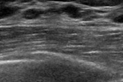
In other news, researchers from Turkey are convinced that an AI model can accurately predict malignancy on breast ultrasound based on BI-RADS assessment. The technique showed comparable performance to that of radiologists and can help avoid unnecessary biopsies and follow-up exams.
A group from the world-famous Karolinska Institute in Stockholm has also presented their findings on breast AI. Don’t miss our report.
In another top story, Italian investigators have reported that contrast-enhanced mammography confirms the effectiveness of ultrasound-guided cryoablation and can serve as a reliable annual follow-up exam.
For a full listing of our recent coverage, please go to the Womens Imaging content area.




![A normal mammogram confirmed by three-year radiologic follow-up illustrates reader-marked regions of interest (ROIs) during (A) unaided (round 1) and (B) artificial intelligence (AI)–assisted (round 2) reading. Each colored dot represents an ROI for recall by a human reader. Readers could mark more than one ROI per case, represented by multiple dots of the same color. During AI-assisted reading, the AI system displayed three visible prompts: two with suspicion of malignancy scores of 35% (left mediolateral oblique [L MLO] and craniocaudal [L CC]) and one with a suspicion of malignancy score of 10% (right craniocaudal [R CC]), shown as polygonal overlays. Without AI, six of 10 readers (60%) marked a false-positive ROI. With AI assistance, this fell to two of 10 (20%). R MLO = right mediolateral oblique.](https://img.auntminnieeurope.com/mindful/smg/workspaces/default/uploads/2026/07/2026-07-14-radiology-mammogram-ai-auto-bias.H0bYO8QlWs.jpg?auto=format%2Ccompress&fit=crop&h=100&q=70&w=100)


![A normal mammogram confirmed by three-year radiologic follow-up illustrates reader-marked regions of interest (ROIs) during (A) unaided (round 1) and (B) artificial intelligence (AI)–assisted (round 2) reading. Each colored dot represents an ROI for recall by a human reader. Readers could mark more than one ROI per case, represented by multiple dots of the same color. During AI-assisted reading, the AI system displayed three visible prompts: two with suspicion of malignancy scores of 35% (left mediolateral oblique [L MLO] and craniocaudal [L CC]) and one with a suspicion of malignancy score of 10% (right craniocaudal [R CC]), shown as polygonal overlays. Without AI, six of 10 readers (60%) marked a false-positive ROI. With AI assistance, this fell to two of 10 (20%). R MLO = right mediolateral oblique.](https://img.auntminnieeurope.com/mindful/smg/workspaces/default/uploads/2026/07/2026-07-14-radiology-mammogram-ai-auto-bias.H0bYO8QlWs.jpg?auto=format%2Ccompress&fit=crop&h=112&q=70&w=112)