
Dear Women's Imaging Insider,
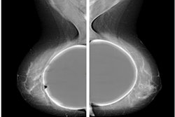
For women with breast implants, tomosynthesis is not currently the technique of choice for screening, but this might soon be changing.
Radiologists in Rome have developed a low-dose protocol that is producing excellent results. We've posted an article today about the team's approach. Find out more in the Women's Imaging Community.
Another recent study that has caught our attention comes from Denmark. The authors explain how AI systems are showing potential for short- and long-term breast cancer risk assessment. Don't miss the commentary from Dr. Filippo Pesapane and a video interview with the lead author.
Any research conducted at the prestigious Karolinska Institute in Stockholm deserves a close look. A paper published in Lancet Digital Health on 8 September found that AI alone and AI with one radiologist are equivalent to double reading by radiologists in breast imaging.
Investigators from the University of Nottingham, U.K., have also reported findings in this area. Check out our news report on their key findings.
The opponents of cancer screening have been relatively quiet of late, but they made a dramatic reappearance last month, when the results of a Norwegian study were published.
The authors don't advocate abandoning cancer screening tests, but they suggest claims of screening saving lives are not supported by the "current best available evidence." They want priorities to be reconsidered, and they think the public should be informed about the benefits, harms, and burden of screening tests.
This letter has highlighted several of the many articles posted over the past month in the Women's Imaging Community. Please take a close look at the full list below, and feel free to contact me if you have ideas for future coverage.








![A normal mammogram confirmed by three-year radiologic follow-up illustrates reader-marked regions of interest (ROIs) during (A) unaided (round 1) and (B) artificial intelligence (AI)–assisted (round 2) reading. Each colored dot represents an ROI for recall by a human reader. Readers could mark more than one ROI per case, represented by multiple dots of the same color. During AI-assisted reading, the AI system displayed three visible prompts: two with suspicion of malignancy scores of 35% (left mediolateral oblique [L MLO] and craniocaudal [L CC]) and one with a suspicion of malignancy score of 10% (right craniocaudal [R CC]), shown as polygonal overlays. Without AI, six of 10 readers (60%) marked a false-positive ROI. With AI assistance, this fell to two of 10 (20%). R MLO = right mediolateral oblique.](https://img.auntminnieeurope.com/mindful/smg/workspaces/default/uploads/2026/07/2026-07-14-radiology-mammogram-ai-auto-bias.H0bYO8QlWs.jpg?auto=format%2Ccompress&dpr=2&fit=crop&h=167&q=70&w=250)


