Dear Women's Imaging Insider,
Prompt and accurate identification of axillary node and internal mammary node metastases in patients with invasive breast cancer is essential to determine the prognosis and decide on the most appropriate treatment.
Authors from Istanbul, Turkey, have shared their experiences of this important area in a new paper posted by the European Journal of Radiology. Don't miss our report, which is today's top article.
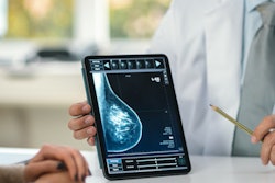
The use of artificial intelligence (AI) in breast imaging was a central theme at the RSNA annual meeting. Among the highlights were research findings from a team in Linköping in Sweden, along with results presented by a group in Central Germany.
Support is also growing for the use of abbreviated breast MRI as a viable alternative to conventional MRI when it comes to high-risk supplemental screening. In another story from RSNA 2022, we've summarized some study results that deserve a close look.
The Chicago congress was a particularly memorable occasion for Dr. Berat Bersu Ozcan, a research fellow in Dallas who received her medical training at Turkey's Hacettepe University. Her digital poster about improving the quality of life of patients through more widely employed breast health initiatives received a prestigious magna cum laude award.
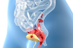
In other news, Italian investigators have described how ultrasound detected signs of endometriosis in approximately one-third of young women presenting with severe dysmenorrhea. Young patients with dysmenorrhea should be referred to an expert sonographer to minimize the delay between the onset of symptoms and diagnosis, the authors stated.
This letter has highlighted some of the numerous articles posted in the Women's Imaging Community over recent weeks. Please take a close look at the full list below, and feel free to contact me if you have ideas for future coverage.




![A normal mammogram confirmed by three-year radiologic follow-up illustrates reader-marked regions of interest (ROIs) during (A) unaided (round 1) and (B) artificial intelligence (AI)–assisted (round 2) reading. Each colored dot represents an ROI for recall by a human reader. Readers could mark more than one ROI per case, represented by multiple dots of the same color. During AI-assisted reading, the AI system displayed three visible prompts: two with suspicion of malignancy scores of 35% (left mediolateral oblique [L MLO] and craniocaudal [L CC]) and one with a suspicion of malignancy score of 10% (right craniocaudal [R CC]), shown as polygonal overlays. Without AI, six of 10 readers (60%) marked a false-positive ROI. With AI assistance, this fell to two of 10 (20%). R MLO = right mediolateral oblique.](https://img.auntminnieeurope.com/mindful/smg/workspaces/default/uploads/2026/07/2026-07-14-radiology-mammogram-ai-auto-bias.H0bYO8QlWs.jpg?auto=format%2Ccompress&fit=crop&h=100&q=70&w=100)


![A normal mammogram confirmed by three-year radiologic follow-up illustrates reader-marked regions of interest (ROIs) during (A) unaided (round 1) and (B) artificial intelligence (AI)–assisted (round 2) reading. Each colored dot represents an ROI for recall by a human reader. Readers could mark more than one ROI per case, represented by multiple dots of the same color. During AI-assisted reading, the AI system displayed three visible prompts: two with suspicion of malignancy scores of 35% (left mediolateral oblique [L MLO] and craniocaudal [L CC]) and one with a suspicion of malignancy score of 10% (right craniocaudal [R CC]), shown as polygonal overlays. Without AI, six of 10 readers (60%) marked a false-positive ROI. With AI assistance, this fell to two of 10 (20%). R MLO = right mediolateral oblique.](https://img.auntminnieeurope.com/mindful/smg/workspaces/default/uploads/2026/07/2026-07-14-radiology-mammogram-ai-auto-bias.H0bYO8QlWs.jpg?auto=format%2Ccompress&fit=crop&h=112&q=70&w=112)