Dear Women's Imaging Insider,
Contrast-enhanced spectral mammography (CESM) -- also known as contrast-enhanced dual-energy mammography -- is making sustained progress and gaining clinical acceptance, but some challenges remain.
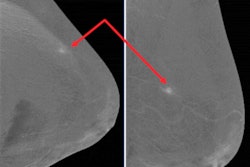
That's the view of Spanish researchers who shared their experiences of CESM recently at the RSNA 2020 meeting. They won a certificate of merit award for their efforts. Don't miss our report posted today.
In other news, forgoing radiation therapy after breast-conserving surgery may not have a negative impact on survival outcomes for some women with breast cancer, according to new study data from Scotland presented at the 2020 San Antonio Breast Cancer Symposium.
A dispute over MRI's role in breast screening is escalating in the Netherlands. The Health Council is against providing MRI screening to women with dense breasts because of the anxiety caused by a false-positive result and unnecessary follow-up diagnostic exams, but the Dutch Society of Radiology is convinced that the evidence confirms the efficacy of screening with MRI in women with dense breasts.
Meanwhile, investigators from the Netherlands and the U.K. have used artificial intelligence (AI) software to pinpoint interval breast cancers. An AI program detected more than one-third of breast cancers missed on screening mammography in two different studies.
Important new statistics about breast screening have emerged from England. A large study has shown a 37% reduction in mortality for women screened at least once, corresponding to about nine breast cancer deaths prevented between the ages of 55 and 79 for every 1,000 women attending screening between the ages of 50 and 69.
These articles make up just part of our Women's Imaging Community reporting. Be sure to check in regularly for current women's imaging news and research developments.



![A normal mammogram confirmed by three-year radiologic follow-up illustrates reader-marked regions of interest (ROIs) during (A) unaided (round 1) and (B) artificial intelligence (AI)–assisted (round 2) reading. Each colored dot represents an ROI for recall by a human reader. Readers could mark more than one ROI per case, represented by multiple dots of the same color. During AI-assisted reading, the AI system displayed three visible prompts: two with suspicion of malignancy scores of 35% (left mediolateral oblique [L MLO] and craniocaudal [L CC]) and one with a suspicion of malignancy score of 10% (right craniocaudal [R CC]), shown as polygonal overlays. Without AI, six of 10 readers (60%) marked a false-positive ROI. With AI assistance, this fell to two of 10 (20%). R MLO = right mediolateral oblique.](https://img.auntminnieeurope.com/mindful/smg/workspaces/default/uploads/2026/07/2026-07-14-radiology-mammogram-ai-auto-bias.H0bYO8QlWs.jpg?auto=format%2Ccompress&fit=crop&h=100&q=70&w=100)


![A normal mammogram confirmed by three-year radiologic follow-up illustrates reader-marked regions of interest (ROIs) during (A) unaided (round 1) and (B) artificial intelligence (AI)–assisted (round 2) reading. Each colored dot represents an ROI for recall by a human reader. Readers could mark more than one ROI per case, represented by multiple dots of the same color. During AI-assisted reading, the AI system displayed three visible prompts: two with suspicion of malignancy scores of 35% (left mediolateral oblique [L MLO] and craniocaudal [L CC]) and one with a suspicion of malignancy score of 10% (right craniocaudal [R CC]), shown as polygonal overlays. Without AI, six of 10 readers (60%) marked a false-positive ROI. With AI assistance, this fell to two of 10 (20%). R MLO = right mediolateral oblique.](https://img.auntminnieeurope.com/mindful/smg/workspaces/default/uploads/2026/07/2026-07-14-radiology-mammogram-ai-auto-bias.H0bYO8QlWs.jpg?auto=format%2Ccompress&fit=crop&h=112&q=70&w=112)