
Dear Women's Imaging Insider,
Digital breast tomosynthesis (DBT) is a much-discussed modality. However, studies often focus on its ability to detect cancer and to reduce recall rates. But how well does it perform for characterizing suspicious lesions? And just how good is synthesized 2D mammography? Find out.
Another DBT study we're highlighting in this edition confirms adding the modality to full-field digital mammography (FFDM) boosts detection rates for invasive cancer, and it may reduce recall and false-positive rates as well. While evidence has suggested DBT performs better than FFDM for diagnostic applications, the relative performance of DBT and FFDM for finding cancer in a screening population has not been fully explored. Until now.
The U.K. continues to experience turmoil after turmoil. First, a contract dispute with the National Health Service resulted in a strike. Now, a new report finds the country's breast screening program is already understaffed and will face worsening personnel problems in the coming years. How bad is it? Read more.
Eye-tracking studies are relatively new and mainly used in pathology, but an Irish study used eye-tracking software for breast imaging. The researchers found radiologists spend significantly more time observing mediolateral oblique breast images and right images. The knowledge may be useful to help remove any interpretation differences and as a feedback tool.
Also in your Women's Imaging Community, breast MRI continues to spark interest. Just how low can radiologists go in terms of protocol times? Pretty low, it turns out. Italian researchers developed an abbreviated breast MRI protocol for characterizing lesions and determined it has the same diagnostic potential as the standard protocol but takes less time.
A study of more than 100,000 women in Egypt has confirmed that an increase in mammographic breast density -- based on the American College of Radiology classifications -- is associated with a heightened risk of breast cancer. Read more.
That's just a small selection of the stories you'll find in your Women's Imaging Community, so head on over there to check out the rest. As always, I love hearing from you so, email me anytime.




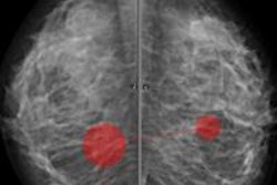




![A normal mammogram confirmed by three-year radiologic follow-up illustrates reader-marked regions of interest (ROIs) during (A) unaided (round 1) and (B) artificial intelligence (AI)–assisted (round 2) reading. Each colored dot represents an ROI for recall by a human reader. Readers could mark more than one ROI per case, represented by multiple dots of the same color. During AI-assisted reading, the AI system displayed three visible prompts: two with suspicion of malignancy scores of 35% (left mediolateral oblique [L MLO] and craniocaudal [L CC]) and one with a suspicion of malignancy score of 10% (right craniocaudal [R CC]), shown as polygonal overlays. Without AI, six of 10 readers (60%) marked a false-positive ROI. With AI assistance, this fell to two of 10 (20%). R MLO = right mediolateral oblique.](https://img.auntminnieeurope.com/mindful/smg/workspaces/default/uploads/2026/07/2026-07-14-radiology-mammogram-ai-auto-bias.H0bYO8QlWs.jpg?auto=format%2Ccompress&dpr=2&fit=crop&h=167&q=70&w=250)


