Dear Women's Imaging Insider,
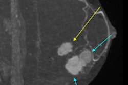
In this month's edition, you'll find many interesting news articles. For instance, we all know breast MRI has high sensitivity, but how does it stack up in a head-to-head comparison with digital breast tomosynthesis (DBT)? MRI and DBT haven't been compared much before, so be sure to check out this new article to see how each modality performed.
Also in your Women's Imaging Community, low- and middle-income countries are seeing a rise in breast cancer incidence and death rates, despite the fact that death rates have decreased in higher-income countries. Although breast cancer was once considered to be primarily a disease of Western women, 52% of new cases and 62% of deaths now occur in economically developing countries. Read more.
German researchers found PET/MRI can provide high diagnostic performance for restaging gynecological cancer patients compared with FDG-PET/CT, with only slightly longer scan times and "markedly reduced" radiation exposure. They found no significant differences between the two hybrid modalities in terms of sensitivity, specificity, positive predictive value, negative predictive value, and diagnostic accuracy.
Ultrasound and x-ray mammography have several limitations for the detection and monitoring of breast cancer, including high false-positive rates and exposure to ionizing radiation. Dutch researchers have started to investigate whether photoacoustic imaging could be a solution. Is it? Find out.
Speaking of problems with mammography, women who receive false-positive mammograms are twice as likely to experience long-term unpleasant psychosocial consequences as women with negative examinations. Swedish researchers found the use of early recall can result in negative emotional consequences that may persist for up to 12 months after the initial false-positive mammogram.
There's more in your Women's Imaging Community, so head on over to read the rest of the articles, or scroll below this message.
As always, I enjoy hearing from you, so keep in touch.