The U.K. National Institute for Health and Care Excellence guidelines indicate that if an incidental liver lesion can be seen on ultrasound, a contrast-enhanced ultrasound (CEUS) exam should be performed as the first-line examination to characterize this lesion. This is often not done in clinical practice, but it is clearly better for the patient and has the benefit of downstream cost savings for healthcare.
Therefore, the need to find out how to make CEUS an integral part of clinical practice is paramount in medicine today. Practitioners must learn how to use CEUS in the kidney, pancreas, spleen, gallbladder, testes, and in many other areas, as well as after interventional liver ablation.
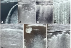
 In a 47-year-old man with upper abdominal pain, ultrasound shows a fatty liver and an atypical lesion (arrow). The CEUS examination answered the question in 30 seconds -- this is a hemangioma, with classical pathognomonic peripheral globular enhancement (arrow). It’s a “slam-dunk” diagnosis: no further imaging, no further costs, and the patient knows immediately that this is benign. Figure courtesy of Prof. Paul Sidhu.
CEUS has become an indispensable “point-of-care” or “problem-solving tool” in clinical practice, allowing for “getting” the answer for a clinical conundrum in minutes, if not seconds. With the current attention of the world media on the cancer-generating potential of excessive CT examinations, we need to show our patients that we are aware of this and have the tools to help avoid unnecessary CT examinations.
With increased patient awareness of the issues of CT radiation and cancer risk, more people will ask for an alternative technique. We should be ready to agree to such requests safely and accurately. We will be put on the spot more and more, so it’s vital to be prepared.
Upcoming EUROSON course
Against this background, the 11th International Contrast Ultrasound Meeting will take place at King’s College Hospital (KCH), London, over two days on 18 and 19 June 2025. The course is supported by the European Federation of Ultrasound Societies in Medicine and Biology (EUROSON), and it will be held at the Windsor Walk Lecture Theatre, Fetal Medicine Research Institute, within easy connection to Central London.
Previous courses have alternated between adult and pediatric applications of CEUS, and this year is the turn of the adults, although some pediatrics is included. The prestigious David Cosgrove Memorial Lecture will be delivered by Prof. Flemming Forsberg, a professor of radiology at Thomas Jefferson University in Philadelphia and an alumnus of KCH, who will demonstrate how you can use CEUS to non-invasively predict portal hypertension accurately -- a game changer for avoiding invasive venous pressures via the hepatic vein wedge pressures.
There is an unprecedented collection of pioneers of CEUS assembled to conduct the course. Previous courses have hit the nail on the head for improving clinical practice with CEUS, and no doubt this one will do the same.
The course directors (Dr. Tim Yusuf, Dr. Annamaria Deganello, and I) guarantee a cost-effective, informative two days with refreshments in abundance (including the course dinner) and a heavily sponsored course fee.
Further details are available on the EFSUMB website. To register, click here.
Prof. Paul Sidhu is professor of imaging sciences at King's College Hospital in London.
The comments and observations expressed herein are those of the authors and do not necessarily reflect the opinions of AuntMinnieEurope.com.