Ultrasound-guided nerve blocks (UGNBs) performed by emergency clinicians appear to be safe for pain management, suggest findings published on 13 November in JAMA Network Open.
Researchers led by Dr. Andrew Goldsmith from Lahey Hospital & Medical Center in Burlington, Massachusetts, U.S., reported that pain experienced by patients who received these blocks was cut at least in half. They also found a small number of complications when implementing UGNBs, which may lead to less reliance on opioids for pain relief.
“Broader implementation of UGNBs in emergency department settings may have important implications as key elements of multimodal analgesia strategies to reduce opioid use and improve patient care,” Goldsmith and co-authors wrote.
UGNBs in recent years have been used for analgesia care in emergency settings to block pain. However, a lack of procedural safety measures has limited their use in this area, the researchers noted. They also highlighted that, to their knowledge, no large-scale multicenter studies evaluating the safety and efficacy of UGNBs by emergency clinicians have been led in the U.S.
The researchers wrote that UGNBs could help lessen reliance on opioids for pain management, given the latter’s negative effects such as misuse, physical dependence, and addiction.
Goldsmith and colleagues studied complication rates stemming from UGNB use in the emergency department and their impact on patient pain scores via a study that included data collected in 2022 and 2023 from 2,735 UGNB encounters among adult patients in 11 U.S. emergency departments. The data came from the National Ultrasound-Guided Nerve Block Registry.
Fascia iliaca blocks, which separate the femoral nerve from the femoral artery, made up 35.6% of the blocks reported. This was the most common UGNB procedure.
Complications occurred in 10 of the total blocks, for a rate of 0.4%. The researchers reported one episode of local anesthetic systemic toxicity that required an intralipid.
Among 1,864 patients who had reported pain levels recorded, 70.8% (n = 1,320) experienced 51% to 100% pain relief following the implementation of UGNBs.
Finally, while operator training level varied, resident physicians performed most UGNB procedures (71.5%).
The study authors suggested that their findings support more widespread adoption of UGNB training and practice across acute care settings, adding that UGNBs present a promising method for multimodal analgesia strategies that could reduce opioid use and improve patient care.
The team called for a large prospective database of UGNB encounters in emergency departments to confirm the findings.
In an accompanying editorial, Dr. Michael Gottlieb from Rush University Medical Center in Chicago, wrote that the study results present an important opportunity to track the use and outcomes of UGNB procedures.
"While the large overall number performed and low rate of complications are promising, the study also highlights the critical need for further efforts to enhance UGNB uptake across sites to ensure adequate skill acquisition and maintenance," he noted.
The study can be found in its entirety here.

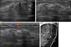
![Examples of ultrasound findings and techniques. (A) Images in a 39-year-old male patient with a mass in the left thigh. The mass is heterogeneous on the B-mode US image (compared with the patient in D) and showed increased microvascularity (superb microvascular imaging [SMI]) and shear-wave elastography (SWE) values. Undifferentiated pleomorphic sarcoma was diagnosed at biopsy (with pleomorphic rhabdomyosarcoma in surgical specimen). (B) Images in an 18-year-old male patient with a mass in the left leg. The mass is hypoechoic on the B-mode image, with no other findings suggestive of malignancy. The lesion is in contact with the cortex of the tibia, which is slightly irregular. CT revealed a doubtful anteromedial tibial erosion. The microvascular study demonstrated high vascularization, suggestive of malignancy. Periosteal Ewing sarcoma was diagnosed with both histologic and immunohistochemical confirmation. (C) Images in a 69-year-old female patient with a lump growing on the outside of the left leg. Multiple SWE examinations were performed (please note the high values obtained in the measurements, whereas the color map highlights the stiffness relative to adjacent tissues). SMI showed areas of increased vascularization to target for sampling. Undifferentiated spindle cell sarcoma was diagnosed at biopsy, with residual leiomyosarcoma in the surgical specimen after neoadjuvant therapy. (D) Images in a 56-year-old female patient with a mass in the right thigh. The mass is heterogeneous at both B-mode ultrasound (similar to patient A) and MRI (coronal T2-weighted spectral attenuated inversion recovery [SPAIR]; T1-weighted pre-contrast and postcontrast imaging), which even shows uptake after the administration of paramagnetic contrast material, which is traditionally suggestive of malignancy. Low values at SMI and elastography are suggestive of benignity. Spindle cell lipoma was diagnosed at biopsy, with atypical spindle cell lipomatous tumor in the surgical specimen.](https://img.auntminnieeurope.com/mindful/smg/workspaces/default/uploads/2026/08/images-radiol250278fig2.APCFLSvX6p.jpg?auto=format%2Ccompress&fit=crop&h=100&q=70&w=100)


![Examples of ultrasound findings and techniques. (A) Images in a 39-year-old male patient with a mass in the left thigh. The mass is heterogeneous on the B-mode US image (compared with the patient in D) and showed increased microvascularity (superb microvascular imaging [SMI]) and shear-wave elastography (SWE) values. Undifferentiated pleomorphic sarcoma was diagnosed at biopsy (with pleomorphic rhabdomyosarcoma in surgical specimen). (B) Images in an 18-year-old male patient with a mass in the left leg. The mass is hypoechoic on the B-mode image, with no other findings suggestive of malignancy. The lesion is in contact with the cortex of the tibia, which is slightly irregular. CT revealed a doubtful anteromedial tibial erosion. The microvascular study demonstrated high vascularization, suggestive of malignancy. Periosteal Ewing sarcoma was diagnosed with both histologic and immunohistochemical confirmation. (C) Images in a 69-year-old female patient with a lump growing on the outside of the left leg. Multiple SWE examinations were performed (please note the high values obtained in the measurements, whereas the color map highlights the stiffness relative to adjacent tissues). SMI showed areas of increased vascularization to target for sampling. Undifferentiated spindle cell sarcoma was diagnosed at biopsy, with residual leiomyosarcoma in the surgical specimen after neoadjuvant therapy. (D) Images in a 56-year-old female patient with a mass in the right thigh. The mass is heterogeneous at both B-mode ultrasound (similar to patient A) and MRI (coronal T2-weighted spectral attenuated inversion recovery [SPAIR]; T1-weighted pre-contrast and postcontrast imaging), which even shows uptake after the administration of paramagnetic contrast material, which is traditionally suggestive of malignancy. Low values at SMI and elastography are suggestive of benignity. Spindle cell lipoma was diagnosed at biopsy, with atypical spindle cell lipomatous tumor in the surgical specimen.](https://img.auntminnieeurope.com/mindful/smg/workspaces/default/uploads/2026/08/images-radiol250278fig2.APCFLSvX6p.jpg?auto=format%2Ccompress&fit=crop&h=112&q=70&w=112)