Dear MRI Insider,
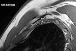
The use of real-time dynamic MRI for evaluating joints relies heavily on suitable patient installation and optimal positioning of the joint in the coil to allow movement, but experts say there is a shortage of guidance for the appropriate protocol, sequence standardizations, and diagnostic criteria for optimum use of MRI.
During last week's Royal Australian and New Zealand College of Radiologists (RANZCR) annual scientific meeting in Adelaide, two radiologists from Brisbane shared their experiences of dynamic imaging at 3 tesla. They explained how advances in MRI techniques and dynamic imaging have helped them to diagnose neurogenic thoracic outlet syndrome. Don't miss our news report posted today.
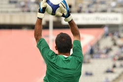
The football World Cup begins on 20 November. Ahead of the tournament, Brazilian researchers have elaborated on the range of hand and wrist injuries incurred by goalkeepers and how they use MRI to improve patient management. Lead author Dr. Tatiane Cantarelli from São Paulo has selected two clinical cases for you.
Lund has a rich heritage when it comes to MRI, and the city hosts Sweden's National 7 Tesla Facility. It's also becoming a focus of attention in the low-field arena, and research is underway into how portable ultralow-field MRI scanners can transform care and improve access. Find out more in the informative article by MRI physicist Emil Ljungberg, PhD.
Advances in MRI-guided percutaneous diagnosis and emerging treatment options for prostate tumors came under scrutiny in a special session held last month at the French national radiology congress, JFR 2022. Top interventional radiologist Prof. Afshin Gangi from Strasbourg was among the speakers.
We've also posted a JFR video interview with Gangi about the structure of French radiology, as well as how to avoid burnout. For French speakers, this interview is also available in a special French-language version. Also, we've now posted a French-language interview with Prof. Denis Le Bihan about diffusion MRI.
In this letter, we've highlighted just a few of the many reports posted in the MRI Community over recent weeks. Please scroll through the full list below, and feel free to contact me if you have ideas for future coverage.


![Overview of the study design. (A) The fully automated deep learning framework was developed to estimate body composition (BC) (defined as subcutaneous adipose tissue [SAT] in liters; visceral adipose tissue [VAT] in liters; skeletal muscle [SM] in liters; SM fat fraction [SMFF] as a percentage; and intramuscular adipose tissue [IMAT] in deciliters) from MRI. The fully automated framework comprised one model (model 1) to quantify different BC measures (SAT, VAT, SM, SMFF, and IMAT) as three-dimensional (3D) measures from whole-body MRI scans. The second model (model 2) was trained to identify standardized anatomic landmarks along the craniocaudal body axis (z coordinate field), which allowed for subdividing the whole-body measures into different subregions typically examined on clinical routine MRI scans (chest, abdomen, and pelvis). (B) BC was quantified from whole-body MRI in over 66,000 individuals from two large population-based cohort studies, the UK Biobank (UKB) (36,317 individuals) and the German National Cohort (NAKO) (30,291 individuals). Bar graphs show age distribution by sex and cohort. BMI = body mass index. (C) After the performance assessment of the fully automated framework, the change in BC measures, distributions, and profiles across age decades were investigated. Age-, sex-, and height-adjusted body composition reference curves were calculated and made publicly available in a web-based z-score calculator (https://circ-ml.github.io).](https://img.auntminnieeurope.com/mindful/smg/workspaces/default/uploads/2026/05/body-comp.XgAjTfPj1W.jpg?auto=format%2Ccompress&fit=crop&h=100&q=70&w=100)


![Overview of the study design. (A) The fully automated deep learning framework was developed to estimate body composition (BC) (defined as subcutaneous adipose tissue [SAT] in liters; visceral adipose tissue [VAT] in liters; skeletal muscle [SM] in liters; SM fat fraction [SMFF] as a percentage; and intramuscular adipose tissue [IMAT] in deciliters) from MRI. The fully automated framework comprised one model (model 1) to quantify different BC measures (SAT, VAT, SM, SMFF, and IMAT) as three-dimensional (3D) measures from whole-body MRI scans. The second model (model 2) was trained to identify standardized anatomic landmarks along the craniocaudal body axis (z coordinate field), which allowed for subdividing the whole-body measures into different subregions typically examined on clinical routine MRI scans (chest, abdomen, and pelvis). (B) BC was quantified from whole-body MRI in over 66,000 individuals from two large population-based cohort studies, the UK Biobank (UKB) (36,317 individuals) and the German National Cohort (NAKO) (30,291 individuals). Bar graphs show age distribution by sex and cohort. BMI = body mass index. (C) After the performance assessment of the fully automated framework, the change in BC measures, distributions, and profiles across age decades were investigated. Age-, sex-, and height-adjusted body composition reference curves were calculated and made publicly available in a web-based z-score calculator (https://circ-ml.github.io).](https://img.auntminnieeurope.com/mindful/smg/workspaces/default/uploads/2026/05/body-comp.XgAjTfPj1W.jpg?auto=format%2Ccompress&fit=crop&h=112&q=70&w=112)


