Dear MRI Insider,
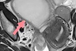
When it comes to endometriosis, MRI rules supreme, and it's particularly important for radiologists to know about the acute complications of endometriomas and to look out for characteristic T1-weighted fat-suppressed foci, say French experts in ob/gyn imaging.
The researchers from CHU Montpellier have shared their experiences of this area, including some impressive clinical images from three cases. Don't miss our news report posted today.
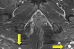
The use of silicone for cosmetic purposes appears to be growing fast, especially in developing countries, according to new work from Latin America. Radiologists must take these procedures into account and learn to recognize the images that they generate, the authors say. MRI can help warn the surgeon about the level of tissue compromise, show the affected planes and the approximate amount of biopolymers injected, and demonstrate their areas in the case of material migration, as shown by three original sets of photos and clinical images.
The London-based radiologist Dr. Simon Rees was a remarkable man. In addition to writing well-regarded textbooks, he helped out at Buckingham Palace, played a key role in treating the injured passengers from an air disaster, and adored the sporting life. Late in his career, he also taught himself about cardiac MRI.
Another U.K. radiologist, Prof. Fiona Gilbert, has vast experience in breast and oncology imaging, and she specializes in MRI. Along with Dr. Erika Denton and Prof. Amaka Offiah, PhD, she's given some practical research tips in a new free podcast.
What will Brexit mean for MRI manufacturers? Market analyst Steve Holloway has addressed this question in a timely article about the new trade deal between the European Union and the U.K. Although some aspects have become clearer, a degree of uncertainty persists, he says.
This letter features only a few of the many reports posted recently in the MRI Community. Please scroll through the full list below, and feel free to contact me if you have ideas for future coverage.


![Overview of the study design. (A) The fully automated deep learning framework was developed to estimate body composition (BC) (defined as subcutaneous adipose tissue [SAT] in liters; visceral adipose tissue [VAT] in liters; skeletal muscle [SM] in liters; SM fat fraction [SMFF] as a percentage; and intramuscular adipose tissue [IMAT] in deciliters) from MRI. The fully automated framework comprised one model (model 1) to quantify different BC measures (SAT, VAT, SM, SMFF, and IMAT) as three-dimensional (3D) measures from whole-body MRI scans. The second model (model 2) was trained to identify standardized anatomic landmarks along the craniocaudal body axis (z coordinate field), which allowed for subdividing the whole-body measures into different subregions typically examined on clinical routine MRI scans (chest, abdomen, and pelvis). (B) BC was quantified from whole-body MRI in over 66,000 individuals from two large population-based cohort studies, the UK Biobank (UKB) (36,317 individuals) and the German National Cohort (NAKO) (30,291 individuals). Bar graphs show age distribution by sex and cohort. BMI = body mass index. (C) After the performance assessment of the fully automated framework, the change in BC measures, distributions, and profiles across age decades were investigated. Age-, sex-, and height-adjusted body composition reference curves were calculated and made publicly available in a web-based z-score calculator (https://circ-ml.github.io).](https://img.auntminnieeurope.com/mindful/smg/workspaces/default/uploads/2026/05/body-comp.XgAjTfPj1W.jpg?auto=format%2Ccompress&fit=crop&h=100&q=70&w=100)


![Overview of the study design. (A) The fully automated deep learning framework was developed to estimate body composition (BC) (defined as subcutaneous adipose tissue [SAT] in liters; visceral adipose tissue [VAT] in liters; skeletal muscle [SM] in liters; SM fat fraction [SMFF] as a percentage; and intramuscular adipose tissue [IMAT] in deciliters) from MRI. The fully automated framework comprised one model (model 1) to quantify different BC measures (SAT, VAT, SM, SMFF, and IMAT) as three-dimensional (3D) measures from whole-body MRI scans. The second model (model 2) was trained to identify standardized anatomic landmarks along the craniocaudal body axis (z coordinate field), which allowed for subdividing the whole-body measures into different subregions typically examined on clinical routine MRI scans (chest, abdomen, and pelvis). (B) BC was quantified from whole-body MRI in over 66,000 individuals from two large population-based cohort studies, the UK Biobank (UKB) (36,317 individuals) and the German National Cohort (NAKO) (30,291 individuals). Bar graphs show age distribution by sex and cohort. BMI = body mass index. (C) After the performance assessment of the fully automated framework, the change in BC measures, distributions, and profiles across age decades were investigated. Age-, sex-, and height-adjusted body composition reference curves were calculated and made publicly available in a web-based z-score calculator (https://circ-ml.github.io).](https://img.auntminnieeurope.com/mindful/smg/workspaces/default/uploads/2026/05/body-comp.XgAjTfPj1W.jpg?auto=format%2Ccompress&fit=crop&h=112&q=70&w=112)


