
Dear AuntMinnieEurope Member,
This week saw the 20th anniversary of arguably the most talked about medical imaging paper of all time: the study of sexual intercourse in an MRI scanner.
The Netherlands is famous for being liberal and progressive, but the Dutch authors still had to conduct their experiments in a totally clandestine manner. Their main aim was to discover whether it was possible to scan the act of coitus as a type of body art. Did they succeed? Find out in our reflective report.
Back in the present, the findings of an important study about the use of artificial intelligence (AI) in breast imaging were published on 17 December.
A team from the world-famous Karolinska Institute in Stockholm found that risk scores provided by their deep-learning model correlated better than breast density-derived risk scores with the patient's diagnosis of cancer within a year. Also, density-based predictors showed decreased performance for more aggressive cancers, whereas the deep learning-based predictor did not. Go to the Women's Imaging Community.
A significant industry story broke this week. Fujifilm is planning to acquire the medical imaging business of Hitachi in what would be a major consolidation of Japanese medical device firms. The 179 billion yen (1.47 billion euro) deal would combine Fujifilm's strength in digital x-ray and image management software with Hitachi's modality scanner business.
Meanwhile, a national survey about MRI safety in Sweden has provided strong evidence that the serious accident in Swedish Lapland on 23 October was not an isolated incident. The authors have called for urgent action to prevent further accidents.
An overhaul of the international classification system for cervical cancer staging took place in 2018, and these changes are having a clinical impact. Researchers from a top London facility have taken a detailed look at the new system, and they won a cum laude award at RSNA 2019 for their work.
This is the final Week in Review of 2019, so may I take this opportunity to send everybody our seasonal greetings from the AuntMinnieEurope.com team. We look forward to bringing you all the latest news in 2020.

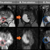


![Overview of the study design. (A) The fully automated deep learning framework was developed to estimate body composition (BC) (defined as subcutaneous adipose tissue [SAT] in liters; visceral adipose tissue [VAT] in liters; skeletal muscle [SM] in liters; SM fat fraction [SMFF] as a percentage; and intramuscular adipose tissue [IMAT] in deciliters) from MRI. The fully automated framework comprised one model (model 1) to quantify different BC measures (SAT, VAT, SM, SMFF, and IMAT) as three-dimensional (3D) measures from whole-body MRI scans. The second model (model 2) was trained to identify standardized anatomic landmarks along the craniocaudal body axis (z coordinate field), which allowed for subdividing the whole-body measures into different subregions typically examined on clinical routine MRI scans (chest, abdomen, and pelvis). (B) BC was quantified from whole-body MRI in over 66,000 individuals from two large population-based cohort studies, the UK Biobank (UKB) (36,317 individuals) and the German National Cohort (NAKO) (30,291 individuals). Bar graphs show age distribution by sex and cohort. BMI = body mass index. (C) After the performance assessment of the fully automated framework, the change in BC measures, distributions, and profiles across age decades were investigated. Age-, sex-, and height-adjusted body composition reference curves were calculated and made publicly available in a web-based z-score calculator (https://circ-ml.github.io).](https://img.auntminnieeurope.com/mindful/smg/workspaces/default/uploads/2026/05/body-comp.XgAjTfPj1W.jpg?auto=format%2Ccompress&dpr=2&fit=crop&h=167&q=70&w=250)