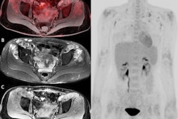
PET/MRI has entered the "Slope of Enlightenment" phase of the Gartner Hype Cycle, according to an editorial published in the November issue of Magnetic Resonance Imaging Clinics of North America.
Prof. Minerva Becker, second vice-president of the European Society of Radiology, and Valentina Garibotto, MD, of University Hospitals of Geneva in Switzerland, introduced a special issue of the journal focusing on advances in PET/MR imaging over the past 10 years. They noted that researchers are now adequately discussing the pros and cons of the approach in critical ways and with evidence-based methodologies.
"The initial hype is over, and the community of PET/MR imaging users is now on the path of enlightenment," they wrote.
Hybrid PET/MRI scanners were introduced in the clinical field in 2010. The approach combines PET molecular imaging data and MRI functional anatomical data in a single patient scan and raised very high expectations early on across almost all clinical applications, the journal editors wrote.
Unsurprisingly, the typical clinical indications for PET/MRI have occurred in the clinical domains in which both PET and MRI were already routinely used as complementary modalities, they noted. Key examples include advanced cancer imaging in different organ systems and metabolic imaging in order to assess the viability of tissue of the brain and heart.
"The main technological progresses focus on quantitative approaches that are of particular interest in an academic environment, given that PET/MR imaging has a place not only in clinical routine but also in research," the authors wrote.
Despite its advantages, the impact of combined PET/MRI on patient care still remains difficult to measure and depends on clinical situations, they added.
Before PET/MRI enters the "Plateau of Productivity" – the final phase of the Hype Cycle – its value must be weighed against the relatively high cost and examination time involved compared with separate acquisitions of each modality. In addition, although the use of multiple biomarkers often facilitates a diagnosis on PET/MRI, this may also be quite challenging, as the different parameters may be contradictory, according to the authors.
"Like many other technical innovations, PET/MR hybrid imaging has followed Gartner’s hype cycle theory that the innovation trigger leads to a peak of inflated expectations, followed by a dip of disillusionment, and finally, a slope of enlightenment leading to a plateau of productivity," they concluded.
The full article is available here.


![Examples of ultrasound findings and techniques. (A) Images in a 39-year-old male patient with a mass in the left thigh. The mass is heterogeneous on the B-mode US image (compared with the patient in D) and showed increased microvascularity (superb microvascular imaging [SMI]) and shear-wave elastography (SWE) values. Undifferentiated pleomorphic sarcoma was diagnosed at biopsy (with pleomorphic rhabdomyosarcoma in surgical specimen). (B) Images in an 18-year-old male patient with a mass in the left leg. The mass is hypoechoic on the B-mode image, with no other findings suggestive of malignancy. The lesion is in contact with the cortex of the tibia, which is slightly irregular. CT revealed a doubtful anteromedial tibial erosion. The microvascular study demonstrated high vascularization, suggestive of malignancy. Periosteal Ewing sarcoma was diagnosed with both histologic and immunohistochemical confirmation. (C) Images in a 69-year-old female patient with a lump growing on the outside of the left leg. Multiple SWE examinations were performed (please note the high values obtained in the measurements, whereas the color map highlights the stiffness relative to adjacent tissues). SMI showed areas of increased vascularization to target for sampling. Undifferentiated spindle cell sarcoma was diagnosed at biopsy, with residual leiomyosarcoma in the surgical specimen after neoadjuvant therapy. (D) Images in a 56-year-old female patient with a mass in the right thigh. The mass is heterogeneous at both B-mode ultrasound (similar to patient A) and MRI (coronal T2-weighted spectral attenuated inversion recovery [SPAIR]; T1-weighted pre-contrast and postcontrast imaging), which even shows uptake after the administration of paramagnetic contrast material, which is traditionally suggestive of malignancy. Low values at SMI and elastography are suggestive of benignity. Spindle cell lipoma was diagnosed at biopsy, with atypical spindle cell lipomatous tumor in the surgical specimen.](https://img.auntminnieeurope.com/mindful/smg/workspaces/default/uploads/2026/08/images-radiol250278fig2.APCFLSvX6p.jpg?auto=format%2Ccompress&fit=crop&h=100&q=70&w=100)


![Examples of ultrasound findings and techniques. (A) Images in a 39-year-old male patient with a mass in the left thigh. The mass is heterogeneous on the B-mode US image (compared with the patient in D) and showed increased microvascularity (superb microvascular imaging [SMI]) and shear-wave elastography (SWE) values. Undifferentiated pleomorphic sarcoma was diagnosed at biopsy (with pleomorphic rhabdomyosarcoma in surgical specimen). (B) Images in an 18-year-old male patient with a mass in the left leg. The mass is hypoechoic on the B-mode image, with no other findings suggestive of malignancy. The lesion is in contact with the cortex of the tibia, which is slightly irregular. CT revealed a doubtful anteromedial tibial erosion. The microvascular study demonstrated high vascularization, suggestive of malignancy. Periosteal Ewing sarcoma was diagnosed with both histologic and immunohistochemical confirmation. (C) Images in a 69-year-old female patient with a lump growing on the outside of the left leg. Multiple SWE examinations were performed (please note the high values obtained in the measurements, whereas the color map highlights the stiffness relative to adjacent tissues). SMI showed areas of increased vascularization to target for sampling. Undifferentiated spindle cell sarcoma was diagnosed at biopsy, with residual leiomyosarcoma in the surgical specimen after neoadjuvant therapy. (D) Images in a 56-year-old female patient with a mass in the right thigh. The mass is heterogeneous at both B-mode ultrasound (similar to patient A) and MRI (coronal T2-weighted spectral attenuated inversion recovery [SPAIR]; T1-weighted pre-contrast and postcontrast imaging), which even shows uptake after the administration of paramagnetic contrast material, which is traditionally suggestive of malignancy. Low values at SMI and elastography are suggestive of benignity. Spindle cell lipoma was diagnosed at biopsy, with atypical spindle cell lipomatous tumor in the surgical specimen.](https://img.auntminnieeurope.com/mindful/smg/workspaces/default/uploads/2026/08/images-radiol250278fig2.APCFLSvX6p.jpg?auto=format%2Ccompress&fit=crop&h=112&q=70&w=112)