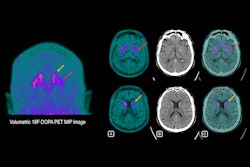
PET brain imaging in individuals with early Parkinson's disease can show which patients are at higher risk of long-term effects, according to a study by researchers from the University of Brescia in Italy.
The team found that hypometabolism patterns on F-18 FDG-PET scans in patients without dementia were associated with worse mental and physical symptoms eight years later. Importantly, these patterns may predict long-term outcomes at the single-patient level, the researchers noted.
"Despite the advances in [Parkinson's disease] clinical subtyping, the identification of patients at higher risk of cognitive and motor progression is still a great challenge at the single-subject level," wrote co-first authors Dr. Alberto Imarisioand Dr Andrea Pilotto and colleagues. The study was published on 7 September in Parkinsonism & Related Disorders.
Clinical presentations are heterogeneous in Parkinson's disease, meaning that symptoms can vary widely, with FDG-PET brain patterns resembling those seen in other neurodegenerative diseases like Dementia with Lewy bodies (DLB) and Alzheimer's disease, the researchers explained.
Moreover, while FDG-PET brain patterns have been previously associated with Parkinson's disease, few studies have explored whether early scans can offer long-term prognostic information, they added.
To that end, the group analyzed imaging from 49 patients with clinical diagnoses of early disease and compared patterns on FDG-PET baseline scans with outcomes in individuals eight years later. They divided the cohort into two groups, those with typical (n = 26) and atypical cortical metabolism (n = 23).
According to the findings, patients with atypical brain hypometabolic patterns showed higher incidence of dementia (60% vs. 3%), hallucinations (56% vs. 7%), and faster motor decline compared with the typical pattern group.
"Importantly, despite presenting brain hypometabolic patterns resembling other neurodegenerative conditions, all the patients included in the atypical pattern group had a confirmed idiopathic [Parkinson's disease] clinical diagnosis," the researchers wrote.
Ultimately, the results expand on the authors' previous work, where they showed an increased risk for dementia at four years in Parkinson's disease patients with Alzheimer's disease-like and DLB-like brain hypometabolic patterns, they wrote. Future work is planned to confirm that FDG-PET scans can differentiate these patients and stratify them based on risk, they noted.
"The present findings show that single-subject brain FDG-PET hypometabolism represents a strong risk factor for long-term cognitive and motor progression in Parkinson's disease," the group concluded.
The full article can be found here.


![Examples of ultrasound findings and techniques. (A) Images in a 39-year-old male patient with a mass in the left thigh. The mass is heterogeneous on the B-mode US image (compared with the patient in D) and showed increased microvascularity (superb microvascular imaging [SMI]) and shear-wave elastography (SWE) values. Undifferentiated pleomorphic sarcoma was diagnosed at biopsy (with pleomorphic rhabdomyosarcoma in surgical specimen). (B) Images in an 18-year-old male patient with a mass in the left leg. The mass is hypoechoic on the B-mode image, with no other findings suggestive of malignancy. The lesion is in contact with the cortex of the tibia, which is slightly irregular. CT revealed a doubtful anteromedial tibial erosion. The microvascular study demonstrated high vascularization, suggestive of malignancy. Periosteal Ewing sarcoma was diagnosed with both histologic and immunohistochemical confirmation. (C) Images in a 69-year-old female patient with a lump growing on the outside of the left leg. Multiple SWE examinations were performed (please note the high values obtained in the measurements, whereas the color map highlights the stiffness relative to adjacent tissues). SMI showed areas of increased vascularization to target for sampling. Undifferentiated spindle cell sarcoma was diagnosed at biopsy, with residual leiomyosarcoma in the surgical specimen after neoadjuvant therapy. (D) Images in a 56-year-old female patient with a mass in the right thigh. The mass is heterogeneous at both B-mode ultrasound (similar to patient A) and MRI (coronal T2-weighted spectral attenuated inversion recovery [SPAIR]; T1-weighted pre-contrast and postcontrast imaging), which even shows uptake after the administration of paramagnetic contrast material, which is traditionally suggestive of malignancy. Low values at SMI and elastography are suggestive of benignity. Spindle cell lipoma was diagnosed at biopsy, with atypical spindle cell lipomatous tumor in the surgical specimen.](https://img.auntminnieeurope.com/mindful/smg/workspaces/default/uploads/2026/08/images-radiol250278fig2.APCFLSvX6p.jpg?auto=format%2Ccompress&fit=crop&h=100&q=70&w=100)


![Examples of ultrasound findings and techniques. (A) Images in a 39-year-old male patient with a mass in the left thigh. The mass is heterogeneous on the B-mode US image (compared with the patient in D) and showed increased microvascularity (superb microvascular imaging [SMI]) and shear-wave elastography (SWE) values. Undifferentiated pleomorphic sarcoma was diagnosed at biopsy (with pleomorphic rhabdomyosarcoma in surgical specimen). (B) Images in an 18-year-old male patient with a mass in the left leg. The mass is hypoechoic on the B-mode image, with no other findings suggestive of malignancy. The lesion is in contact with the cortex of the tibia, which is slightly irregular. CT revealed a doubtful anteromedial tibial erosion. The microvascular study demonstrated high vascularization, suggestive of malignancy. Periosteal Ewing sarcoma was diagnosed with both histologic and immunohistochemical confirmation. (C) Images in a 69-year-old female patient with a lump growing on the outside of the left leg. Multiple SWE examinations were performed (please note the high values obtained in the measurements, whereas the color map highlights the stiffness relative to adjacent tissues). SMI showed areas of increased vascularization to target for sampling. Undifferentiated spindle cell sarcoma was diagnosed at biopsy, with residual leiomyosarcoma in the surgical specimen after neoadjuvant therapy. (D) Images in a 56-year-old female patient with a mass in the right thigh. The mass is heterogeneous at both B-mode ultrasound (similar to patient A) and MRI (coronal T2-weighted spectral attenuated inversion recovery [SPAIR]; T1-weighted pre-contrast and postcontrast imaging), which even shows uptake after the administration of paramagnetic contrast material, which is traditionally suggestive of malignancy. Low values at SMI and elastography are suggestive of benignity. Spindle cell lipoma was diagnosed at biopsy, with atypical spindle cell lipomatous tumor in the surgical specimen.](https://img.auntminnieeurope.com/mindful/smg/workspaces/default/uploads/2026/08/images-radiol250278fig2.APCFLSvX6p.jpg?auto=format%2Ccompress&fit=crop&h=112&q=70&w=112)