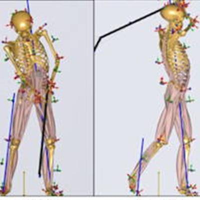
Researchers have visualized golf swing kinetics in patients who have had hip replacements and found that the swing does not contribute excessive force to the implant, according to a study published on 29 May in Scientific Reports.
A group led by orthopedic surgeon Tetsunari Harada, MD, of Kyushu University in Fukuoka used image-matching techniques to study 3D hip ranges of motion (RoMs) and hip contact force in a group of recreational golfers following total hip arthroplasty (THA). Using a driver to "tee off" exerts about as much force on the implants as jogging, they found.
"While golf is not considered to be a high-impact sport and the majority of attending surgeons recommend a return to golf after THA, no previous study has addressed the kinetic data of golfers performing a drive after THA," the group wrote.
To that end, the researchers recruited 10 primary THA patients from their hospital to participate in a study of their driver golf swing. They used a 10-camera Vicon motion capture system paired to a musculoskeletal modeling system scaled to each patient's body size. Input data included leg length differences and femoral offset acquired on x-ray for each patient before and after surgery.
Patient's (recreational right-handed golfer) driver golf swing. (A) Actual driver swing scene, (B) Motion capture system (VICON), (C) Musculoskeletal modeling system (AnyBody). Image and caption courtesy of Scientific Reports.During a driver swing with a mean club head speed of 31 meters per second, implanted hip rotations were between 20 to 30 degrees -- equivalent to those in normal hips analyzed by the same motion capture methods, according to the findings.
The mean hip contact force for the lead replaced hip (depending on the golfer's stance) was 5.5 times patient body weight and 6.7 times body weight for the trail replaced hips, with a tendency of larger force on trail hips in both left THA and right THA, according to the results. This is somewhat lower than the hip contact force exerted on implants while jogging at 6 to 8 km/h, according to the group.
"The number of golf swing motions is much less than the number of steps taken while jogging and the repetition of loading cycles is significantly smaller," the authors noted.
Ultimately, excessive force can have negative effects on implants and lead to complications, including implant failure, the authors noted. This is the first report on hip kinetics including contact forces during the golf swing after THA, they added.
Limitations included only analyzing swings using a driver versus other clubs and that participants performed while barefoot and without a golf ball, which could have altered swing mechanics, the researchers wrote.
"The driver swing of unilateral THA patients did not contribute excessive hip contact forces to the right and left hip prosthesis," the group concluded.
The full article can be found on Scientific Reports.


![Examples of ultrasound findings and techniques. (A) Images in a 39-year-old male patient with a mass in the left thigh. The mass is heterogeneous on the B-mode US image (compared with the patient in D) and showed increased microvascularity (superb microvascular imaging [SMI]) and shear-wave elastography (SWE) values. Undifferentiated pleomorphic sarcoma was diagnosed at biopsy (with pleomorphic rhabdomyosarcoma in surgical specimen). (B) Images in an 18-year-old male patient with a mass in the left leg. The mass is hypoechoic on the B-mode image, with no other findings suggestive of malignancy. The lesion is in contact with the cortex of the tibia, which is slightly irregular. CT revealed a doubtful anteromedial tibial erosion. The microvascular study demonstrated high vascularization, suggestive of malignancy. Periosteal Ewing sarcoma was diagnosed with both histologic and immunohistochemical confirmation. (C) Images in a 69-year-old female patient with a lump growing on the outside of the left leg. Multiple SWE examinations were performed (please note the high values obtained in the measurements, whereas the color map highlights the stiffness relative to adjacent tissues). SMI showed areas of increased vascularization to target for sampling. Undifferentiated spindle cell sarcoma was diagnosed at biopsy, with residual leiomyosarcoma in the surgical specimen after neoadjuvant therapy. (D) Images in a 56-year-old female patient with a mass in the right thigh. The mass is heterogeneous at both B-mode ultrasound (similar to patient A) and MRI (coronal T2-weighted spectral attenuated inversion recovery [SPAIR]; T1-weighted pre-contrast and postcontrast imaging), which even shows uptake after the administration of paramagnetic contrast material, which is traditionally suggestive of malignancy. Low values at SMI and elastography are suggestive of benignity. Spindle cell lipoma was diagnosed at biopsy, with atypical spindle cell lipomatous tumor in the surgical specimen.](https://img.auntminnieeurope.com/mindful/smg/workspaces/default/uploads/2026/08/images-radiol250278fig2.APCFLSvX6p.jpg?auto=format%2Ccompress&fit=crop&h=100&q=70&w=100)


![Examples of ultrasound findings and techniques. (A) Images in a 39-year-old male patient with a mass in the left thigh. The mass is heterogeneous on the B-mode US image (compared with the patient in D) and showed increased microvascularity (superb microvascular imaging [SMI]) and shear-wave elastography (SWE) values. Undifferentiated pleomorphic sarcoma was diagnosed at biopsy (with pleomorphic rhabdomyosarcoma in surgical specimen). (B) Images in an 18-year-old male patient with a mass in the left leg. The mass is hypoechoic on the B-mode image, with no other findings suggestive of malignancy. The lesion is in contact with the cortex of the tibia, which is slightly irregular. CT revealed a doubtful anteromedial tibial erosion. The microvascular study demonstrated high vascularization, suggestive of malignancy. Periosteal Ewing sarcoma was diagnosed with both histologic and immunohistochemical confirmation. (C) Images in a 69-year-old female patient with a lump growing on the outside of the left leg. Multiple SWE examinations were performed (please note the high values obtained in the measurements, whereas the color map highlights the stiffness relative to adjacent tissues). SMI showed areas of increased vascularization to target for sampling. Undifferentiated spindle cell sarcoma was diagnosed at biopsy, with residual leiomyosarcoma in the surgical specimen after neoadjuvant therapy. (D) Images in a 56-year-old female patient with a mass in the right thigh. The mass is heterogeneous at both B-mode ultrasound (similar to patient A) and MRI (coronal T2-weighted spectral attenuated inversion recovery [SPAIR]; T1-weighted pre-contrast and postcontrast imaging), which even shows uptake after the administration of paramagnetic contrast material, which is traditionally suggestive of malignancy. Low values at SMI and elastography are suggestive of benignity. Spindle cell lipoma was diagnosed at biopsy, with atypical spindle cell lipomatous tumor in the surgical specimen.](https://img.auntminnieeurope.com/mindful/smg/workspaces/default/uploads/2026/08/images-radiol250278fig2.APCFLSvX6p.jpg?auto=format%2Ccompress&fit=crop&h=112&q=70&w=112)