TORONTO - Magnetic particle imaging (MPI) with new iron oxide-based contrast agents shows promise for visualizing neuroinflammation, according to a presentation at the International Society for Magnetic Resonance in Medicine (ISMRM) meeting.
In a basic science session on neuroinflammation on 4 June, Maxime Gauberti, MD, of Normandie University in Caen, France, presented preclinical research involving imaging of endothelial cell activation in mice to reveal inflammatory processes in the brain. The research presents new developments decades in the making, he said.
"The ability to detect a molecular target in the central nervous system noninvasively and at high spatial resolution using MRI has attracted the interest of researchers for several decades," he said.
Neuroinflammation is a key immune response observed in many neurological and neurodegenerative diseases. Currently, MRI of neuroinflammation relies on two main imaging features -- edema and blood-brain barrier leakage -- yet these features are not specific for inflammation and the diagnosis of inflammatory lesions of the central nervous system remains a challenge, Gauberti explained.
One problem is that current contrast agents such as gadolinium do not cross the brain's protective blood-brain barrier unless there is leakage due to injury, for instance. Moreover, particles must be smaller than 50 nanometers to escape being cleared from the brain by the body's immune system and they need to be present in high enough amounts to be detected by imaging, he said.
To solve these issues, Gauberti and colleagues are targeting a cellular process called leukocyte-endothelial cell adhesion, which is triggered by injury to brain tissue. During the process, activated leukocytes migrate from the bloodstream to the sites of injury and drive inflammation, which destroys healthy cells.
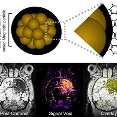
Mapping neuroinflammation using a new family of contrast agents ("Immuno-MRI") targeting activated endothelial cells in the central nervous system. The clinical development of this new tool is ongoing. Image courtesy of Dr. Maxime Gauberti.To visualize this process, the researchers synthesized microparticles of iron oxide (MPIO) that are small enough to cross the blood-brain barrier and that bind to these leukocyte-endothelial cell interactions. Once injected, the MPIO agents bind to the sites where they are detected on imaging.
In studies in mice, the so-called "Immuno-MRI" technique looks promising, and it has revealed neuroinflammation compared to normal subjects, Gauberti said.
Ultimately, one goal of these preclinical studies is to eventually combine conventional MRI with these molecular imaging techniques to aid in the diagnosis of diseases associated with neuroinflammation, such as Alzheimer's disease.
Gauberti added that the research has recently received a boost with the development of specific scanners dedicated to magnetic particle imaging, with one installed recently at his group's center in Caen.
"We can expect in the future the development of magnetic particle imaging as a new modality to look at more neuroinflammation information with these techniques," Gauberti concluded.


![Examples of ultrasound findings and techniques. (A) Images in a 39-year-old male patient with a mass in the left thigh. The mass is heterogeneous on the B-mode US image (compared with the patient in D) and showed increased microvascularity (superb microvascular imaging [SMI]) and shear-wave elastography (SWE) values. Undifferentiated pleomorphic sarcoma was diagnosed at biopsy (with pleomorphic rhabdomyosarcoma in surgical specimen). (B) Images in an 18-year-old male patient with a mass in the left leg. The mass is hypoechoic on the B-mode image, with no other findings suggestive of malignancy. The lesion is in contact with the cortex of the tibia, which is slightly irregular. CT revealed a doubtful anteromedial tibial erosion. The microvascular study demonstrated high vascularization, suggestive of malignancy. Periosteal Ewing sarcoma was diagnosed with both histologic and immunohistochemical confirmation. (C) Images in a 69-year-old female patient with a lump growing on the outside of the left leg. Multiple SWE examinations were performed (please note the high values obtained in the measurements, whereas the color map highlights the stiffness relative to adjacent tissues). SMI showed areas of increased vascularization to target for sampling. Undifferentiated spindle cell sarcoma was diagnosed at biopsy, with residual leiomyosarcoma in the surgical specimen after neoadjuvant therapy. (D) Images in a 56-year-old female patient with a mass in the right thigh. The mass is heterogeneous at both B-mode ultrasound (similar to patient A) and MRI (coronal T2-weighted spectral attenuated inversion recovery [SPAIR]; T1-weighted pre-contrast and postcontrast imaging), which even shows uptake after the administration of paramagnetic contrast material, which is traditionally suggestive of malignancy. Low values at SMI and elastography are suggestive of benignity. Spindle cell lipoma was diagnosed at biopsy, with atypical spindle cell lipomatous tumor in the surgical specimen.](https://img.auntminnieeurope.com/mindful/smg/workspaces/default/uploads/2026/08/images-radiol250278fig2.APCFLSvX6p.jpg?auto=format%2Ccompress&fit=crop&h=100&q=70&w=100)


![Examples of ultrasound findings and techniques. (A) Images in a 39-year-old male patient with a mass in the left thigh. The mass is heterogeneous on the B-mode US image (compared with the patient in D) and showed increased microvascularity (superb microvascular imaging [SMI]) and shear-wave elastography (SWE) values. Undifferentiated pleomorphic sarcoma was diagnosed at biopsy (with pleomorphic rhabdomyosarcoma in surgical specimen). (B) Images in an 18-year-old male patient with a mass in the left leg. The mass is hypoechoic on the B-mode image, with no other findings suggestive of malignancy. The lesion is in contact with the cortex of the tibia, which is slightly irregular. CT revealed a doubtful anteromedial tibial erosion. The microvascular study demonstrated high vascularization, suggestive of malignancy. Periosteal Ewing sarcoma was diagnosed with both histologic and immunohistochemical confirmation. (C) Images in a 69-year-old female patient with a lump growing on the outside of the left leg. Multiple SWE examinations were performed (please note the high values obtained in the measurements, whereas the color map highlights the stiffness relative to adjacent tissues). SMI showed areas of increased vascularization to target for sampling. Undifferentiated spindle cell sarcoma was diagnosed at biopsy, with residual leiomyosarcoma in the surgical specimen after neoadjuvant therapy. (D) Images in a 56-year-old female patient with a mass in the right thigh. The mass is heterogeneous at both B-mode ultrasound (similar to patient A) and MRI (coronal T2-weighted spectral attenuated inversion recovery [SPAIR]; T1-weighted pre-contrast and postcontrast imaging), which even shows uptake after the administration of paramagnetic contrast material, which is traditionally suggestive of malignancy. Low values at SMI and elastography are suggestive of benignity. Spindle cell lipoma was diagnosed at biopsy, with atypical spindle cell lipomatous tumor in the surgical specimen.](https://img.auntminnieeurope.com/mindful/smg/workspaces/default/uploads/2026/08/images-radiol250278fig2.APCFLSvX6p.jpg?auto=format%2Ccompress&fit=crop&h=112&q=70&w=112)