Musculoskeletal (MSK) radiologist Dr. Tom Entwisle has dodged a jail sentence after driving a golf buggy through the central business district (CBD) of Adelaide while under the influence of alcohol, according to reports in the Australian media.
Entwisle, 44, pleaded guilty to driving the vehicle with a blood-alcohol level of 0.132%, the Australian Broadcasting Corporation (ABC) reported on 18 May. A level of 0.08% is widely recognized to be the legal limit for "driving under the influence" of alcohol.
The incident happened during the WOMADelaide, an annual four-day music, arts, and dance festival. Police apprehended Entwisle in the early hours of 11 March, after he had attended the festival, ABC noted.
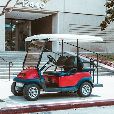
Entwisle had been drinking at the event throughout the day and evening before he and some friends started helping the staff to clean up. "You and one of your friends were using a WOMAD golf buggy to move chairs at this time," said Magistrate John Wells. "Unfortunately, your friend took off in the golf buggy, driving away from Botanic Park and towards the city."
Wells said Entwisle made the "foolish decision" to take over control of the vehicle before police caught up with him. "There is CCTV of your driving. You were driving slowly with minimal pedestrians around."
Given the nature of the vehicle and the lack of other road users or pedestrians, Wells said he was prepared to conclude there was a "less pronounced" risk to the community. The court heard that Entwisle was deeply ashamed of his actions and pleaded guilty to the offense at the first available opportunity, ABC continued. The magistrate acknowledged that it was unusual to deal with drink-driving matters without conviction, but he was satisfied the radiologist was unlikely to re-offend.
"This experience, standing in the dock of a criminal court, the associated shame and public scrutiny all cohere into a strong motivation for you not to re-offend," Wells said. "You have been exposed to publicity and additional embarrassment that most people who plead guilty to this offence do not."
Another article (for subscribers only) posted on 18 May by The Advertiser, a tabloid newspaper in Adelaide, used the headline: "Top doctor in drunken CBD golf buggy joyride."
Entwisle was given a community work order and is disqualified from driving for nine months, backdated to March, the ABC article concluded.
Entwisle is involved in academic research in all areas of MSK imaging, but his main interests involve muscle injury, according to his bio on the Fowler Simmons Radiology website. He completed his postgraduate training in radiology at Austin Health in Melbourne, where there was an emphasis on intervention and hepatobiliary, spinal, neurology, oncology, and emergency imaging. Entwisle started at Imaging @ Olympic Park as a fellow in 2015 and continued as a consultant in 2016, the bio stated.


![Examples of ultrasound findings and techniques. (A) Images in a 39-year-old male patient with a mass in the left thigh. The mass is heterogeneous on the B-mode US image (compared with the patient in D) and showed increased microvascularity (superb microvascular imaging [SMI]) and shear-wave elastography (SWE) values. Undifferentiated pleomorphic sarcoma was diagnosed at biopsy (with pleomorphic rhabdomyosarcoma in surgical specimen). (B) Images in an 18-year-old male patient with a mass in the left leg. The mass is hypoechoic on the B-mode image, with no other findings suggestive of malignancy. The lesion is in contact with the cortex of the tibia, which is slightly irregular. CT revealed a doubtful anteromedial tibial erosion. The microvascular study demonstrated high vascularization, suggestive of malignancy. Periosteal Ewing sarcoma was diagnosed with both histologic and immunohistochemical confirmation. (C) Images in a 69-year-old female patient with a lump growing on the outside of the left leg. Multiple SWE examinations were performed (please note the high values obtained in the measurements, whereas the color map highlights the stiffness relative to adjacent tissues). SMI showed areas of increased vascularization to target for sampling. Undifferentiated spindle cell sarcoma was diagnosed at biopsy, with residual leiomyosarcoma in the surgical specimen after neoadjuvant therapy. (D) Images in a 56-year-old female patient with a mass in the right thigh. The mass is heterogeneous at both B-mode ultrasound (similar to patient A) and MRI (coronal T2-weighted spectral attenuated inversion recovery [SPAIR]; T1-weighted pre-contrast and postcontrast imaging), which even shows uptake after the administration of paramagnetic contrast material, which is traditionally suggestive of malignancy. Low values at SMI and elastography are suggestive of benignity. Spindle cell lipoma was diagnosed at biopsy, with atypical spindle cell lipomatous tumor in the surgical specimen.](https://img.auntminnieeurope.com/mindful/smg/workspaces/default/uploads/2026/08/images-radiol250278fig2.APCFLSvX6p.jpg?auto=format%2Ccompress&fit=crop&h=100&q=70&w=100)


![Examples of ultrasound findings and techniques. (A) Images in a 39-year-old male patient with a mass in the left thigh. The mass is heterogeneous on the B-mode US image (compared with the patient in D) and showed increased microvascularity (superb microvascular imaging [SMI]) and shear-wave elastography (SWE) values. Undifferentiated pleomorphic sarcoma was diagnosed at biopsy (with pleomorphic rhabdomyosarcoma in surgical specimen). (B) Images in an 18-year-old male patient with a mass in the left leg. The mass is hypoechoic on the B-mode image, with no other findings suggestive of malignancy. The lesion is in contact with the cortex of the tibia, which is slightly irregular. CT revealed a doubtful anteromedial tibial erosion. The microvascular study demonstrated high vascularization, suggestive of malignancy. Periosteal Ewing sarcoma was diagnosed with both histologic and immunohistochemical confirmation. (C) Images in a 69-year-old female patient with a lump growing on the outside of the left leg. Multiple SWE examinations were performed (please note the high values obtained in the measurements, whereas the color map highlights the stiffness relative to adjacent tissues). SMI showed areas of increased vascularization to target for sampling. Undifferentiated spindle cell sarcoma was diagnosed at biopsy, with residual leiomyosarcoma in the surgical specimen after neoadjuvant therapy. (D) Images in a 56-year-old female patient with a mass in the right thigh. The mass is heterogeneous at both B-mode ultrasound (similar to patient A) and MRI (coronal T2-weighted spectral attenuated inversion recovery [SPAIR]; T1-weighted pre-contrast and postcontrast imaging), which even shows uptake after the administration of paramagnetic contrast material, which is traditionally suggestive of malignancy. Low values at SMI and elastography are suggestive of benignity. Spindle cell lipoma was diagnosed at biopsy, with atypical spindle cell lipomatous tumor in the surgical specimen.](https://img.auntminnieeurope.com/mindful/smg/workspaces/default/uploads/2026/08/images-radiol250278fig2.APCFLSvX6p.jpg?auto=format%2Ccompress&fit=crop&h=112&q=70&w=112)